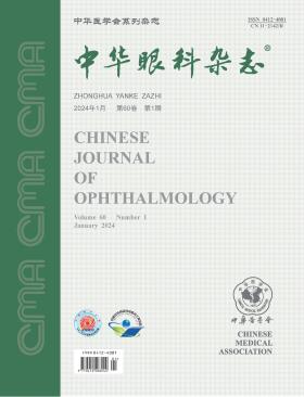
Chinese Journal of Ophthalmology
Volume 60 · Issue 01 · 2024
Chin J Ophthalmol
Farewell to a fruitful 2023, the Chinese Journal of Ophthalmology ushered in a new chapter of Volume 60. Editor-in-chief Yao Ke pointed out in the article "Talking about Promoting the All-round Development of China's Eye Health with Innovation as the Core" in the foreword of this issue that in the field of ophthalmology, innovation is the main indicator to measure whether basic research and clinical research in ophthalmology are advanced. Practicing the innovation-driven development strategy in actual work is the focus and main direction of China's ophthalmology in the future. All ophthalmologists should strengthen their confidence, seize the opportunity, take advantage of the situation, make new contributions to ophthalmology technological innovation, and contribute new strength to the all-round development of China's eye health cause.
In 2023, * emphasized that we should adhere to the "four orientations", accelerate the implementation of the innovation-driven development strategy, promote in-depth cooperation between industry, university and research, focus on strengthening the construction of major scientific and technological innovation platforms, support top scientists to take the lead in original and leading scientific and technological research, strive to break through key core technical problems, and achieve independent controllability in key areas and key links. Innovation is the eternal theme of scientific and technological development. In the future, the competition in the world will mainly focus on scientific and technological innovation, which requires us to think seriously and actively prepare. In the field of ophthalmology, innovation is the main indicator to measure whether basic and clinical research in ophthalmology are advanced. Practicing the innovation-driven development strategy in actual work is the focus and main direction of China's ophthalmology in the future. Combined with the current situation at home and abroad and the present situation of ophthalmology development, it is suggested to carry out the work from the following five aspects.
Superior oblique muscle paralysis is a common type of vertical rotatory strabismus with various subtypes. Regardless of the subtype, the Bielschowsky tilt test plays a crucial role in the diagnosis of superior oblique muscle paralysis and is often considered a significant criterion for diagnosis and differential diagnosis. However, the sensitivity and specificity of the Bielschowsky tilt test for diagnosing superior oblique muscle paralysis are not 100% due to the mechanism involved. The test is not solely based on extraocular muscle imbalance but also involves reflex pathways of the vestibular system and central nervous system. Consequently, lesions affecting corresponding areas may yield positive results in the Bielschowsky tilt test. Additionally, vestibular and central nervous system lesions can also cause strabismus, leading to a lack of one-to-one correspondence between a positive Bielschowsky tilt test and superior oblique muscle paralysis. Therefore, correctly interpreting the role of the Bielschowsky tilt test in superior oblique muscle paralysis is of paramount importance for the effective clinical management and treatment of associated conditions.
Childhood vision is in the stage of development, and the incidence of ametropia is high. For special types of refractive abnormalities such as ultra-high myopia, ultra-high anisometropia and aphakia, conventional wearing of frame glasses can easily cause aberrations and peripheral visual deformations, affect the visual development of children, and even cause refractory amblyopia. In this article, the benefits of contact lenses are discussed from perspectives of ultra-high refractive error, refraction-related refractory amblyopia, and photosensitivity-related refractive error, so as to attract the attention of clinical physicians, expand the application scope of contact lens wear, give full play to the role of contact lenses, and improve the diagnosis and treatment of refractive abnormalities in children.
Myopia in children shows a trend of high incidence and younger age. To standardize the integrated Chinese and western medicine methods of diagnosis, classification, prevention and control of myopia in children, as well as to improve the prevention and control effectiveness, the Chinese Association of Integration Medicine, China Association of Chinese Medicine and Chinese Medical Association have gathered relevant experts to investigate clinical problems, retrieve, screen, extract and synthesize evidence, and form expert consensus opinions in accordance with the requirements of evidence-based medicine methodology. The diagnosis and treatment guideline for integrated Chinese and western medicine for myopia prevention and control in children includes recommendations on how to effectively delay the occurrence of myopia in healthy children, avoid the development of pseudomyopia to myopia, delay the development of low myopia to high myopia, prevent and treat complications of high myopia. Eye health-related behavior monitoring and correction, acupuncture, transcutaneous electrical acupoint stimulation, optics, and medications are introduced. This guideline can provide guidance not only for medical professionals and technical personnel in clinical and scientific research practice, but also for the myopic, pseudomyopic, premyopic and healthy population in preventing and controlling myopia.
To investigate the clinical features, imaging manifestations, histopathological characteristics, and surgical outcomes in pediatric progressive restrictive strabismus.
A retrospective case series study was conducted, including data from 9 cases (9 eyes) of pediatric progressive restrictive strabismus treated at Tianjin Eye Hospital from June 2017 to October 2022. The study compared the degree of globe protrusion in both eyes, changes in eyelid fissure height during internal and external rotation in the primary gaze, summarized clinical characteristics, and analyzed intraoperative conditions, surgical outcomes and postoperative histopathological results of strabismus correction surgery. Statistical analysis was performed using Wilcoxon signed-rank test and Friedman two-way analysis of variance.
All 9 cases involved unilateral onset, with 4 males and 5 females. Three cases affected the right eye, and six affected the left eye. Onset age ranged from 2 to 40 months. The degree of globe protrusion in the affected eyes was 13.00 (12.00, 13.00) mm for the right eye and 12.00 (12.00, 13.50) mm for the left eye, with no statistically significant difference (Z=-1.00, P=0.317). There were no significant changes in eyelid fissure height during internal rotation [8.00 (7.25, 8.00) mm], primary gaze [7.50 (7.00, 8.00) mm], and external rotation [8.00 (7.75, 8.00) mm] in the affected eyes (χ²=1.00, P=0.607). No apparent abnormalities were observed in head CT or MRI scans, serum, or immunological tests. However, orbital CT or MRI scans indicated thickening of different extraocular muscle bellies. Six out of nine cases underwent strabismus correction surgery, and postoperative examination revealed restriction in eye movement despite achieving orthophoria in the primary gaze. Tissue pathology of three cases showed increased collagen fiber proliferation in one, scattered bundles of smooth muscle fibers amid diffuse collagen fiber proliferation in another, and abnormal proliferation of striated muscle fibers with varying diameters, increased paired box (PAX)7-positive satellite cells expressing slow muscle myosin in the third case.
Pediatric progressive restrictive strabismus presents with restrictive changes, without significant alterations in globe protrusion and eyelid fissure height. Imaging examinations reveal thickening of the extraocular muscle bellies in the affected eye. Although strabismus correction surgery improves eye position, postoperative eye movement remains restricted. Histopathological findings in some cases show abnormal proliferation of skeletal muscle fibers or collagen fibers.
To describe clinical characteristics and surgical outcomes of pediatric epiretinal membranes (ERMs) without specific etiologies.
Medical data of a cohort of pediatric patients (≤14 years) who had ERMs without specific etiologies, underwent surgical removal from January 2019 to September 2021, and were followed up for at least 12 months were retrospectively reviewed. Age at presentation, chief complaints, color fundus photographs, optical coherence tomographic images, preoperative and postoperative visual acuities, anatomical changes, and postoperative complications were assessed.
There were 14 patients (17 eyes), including 5 females (6 eyes) and 9 males (11 eyes). The mean age at surgery was 6.31±2.91 years, and the follow-up duration was 17.3±9.5 months. Eight patients were found to have low vision in the school physical examination. Fifteen eyes had an appearance of cellophane macular reflex on fundus images. On optical coherence tomographic images, 10 eyes had"taco"folds, and 7 eyes had"ripple"folds. Five eyes had ellipsoid zone disruptions, while 12 eyes had ellipsoid zone integrity. The preoperative and postoperative best-corrected visual acuities in logMAR were 0.532±0.302 and 0.340±0.298. One patient suffered traumatic cataract and secondary retinal detachment postoperatively, and after further vitrectomy, the retina became attached.
Pediatric ERMs without specific etiologies were mostly found in school-age children with cellophane macular reflex and"taco"folds. Vitrectomy may result in both potential visual acuity and macular anatomical improvements.
To observe the efficacy and safety of botulinum toxin A (BTA) injection in the treatment of acute comitant esotropia (ACE) with different doses.
This retrospective cohort study included data from patients with ACE who received BTA injection treatment at the First Affiliated Hospital of Zhengzhou University from June 2019 to June 2022. All patients underwent routine ophthalmic examinations, including best-corrected visual acuity (BCVA), spherical equivalent (SE), as well as specialized examinations for strabismus, including the degree of esotropia, eye movement status, and binocular visual function. Patients were categorized into small esotropia [≤60 prism diopters (PD)] and large esotropia (>60 PD) groups based on the pre-treatment degree of esotropia. Each group was further divided into 2.5 U and 5.0 U dose subgroups. Monocular injections were administered to the non-dominant eye. The esotropia degree was recorded and compared at 1, 2, 3, and 6 months of follow-up. The proportion of effectively treated patients in each group was documented. The number of cases with various levels of visual functions (including simultaneous vision, near stereopsis, and distance stereopsis) at 6 months post-treatment was compared, and complications during the follow-up period were observed. Statistical analyses were conducted using t-tests, Mann-Whitney U tests, and χ2 tests.
A total of 70 patients were included in the study, comprising 46 males and 24 females, with a median age of 5.0 (4.0, 8.3) years. Among them, 37 patients had small esotropia, with 25 in the 2.5 U group and 12 in the 5.0 U group. Thirty-three patients had large esotropia, with 18 in the 2.5 U group and 15 in the 5.0 U group. There were no statistically significant differences in baseline data, including age, duration of the condition, pre-treatment esotropia degree, BCVA and SE, between the two dose groups in both small and large esotropia patients (all P>0.05). In small esotropia patients, at 1 and 2 months post-treatment, the esotropia degree in the 5.0 U group was -20.00 (-37.50, -7.00) and 0.00 (0.00, 0.00) PD, respectively, which was significantly lower than the 0.00 (-10.00, 4.50) and 5.00 (0.00, 6.50) PD in the 2.5 U group (all P<0.05). At 3 and 6 months post-treatment, the esotropia degree in the 2.5 U group was 5.00 (0.00, 15.00) and 2.00 (0.00, 6.00) PD, respectively, while in the 5.0 U group, it was 0.00 (0.00, 4.50) and 0.00 (0.00, 3.75) PD, with no statistically significant differences between the two groups (all P>0.05). In the 2.5 U group, 20 cases were effectively treated, accounting for 80.0%, while in the 5.0 U group, 10 cases were effective, accounting for 10/12, with no significant difference between the two groups (P>0.05). In the 2.5 U group and the 5.0 U group, the proportions of cases with various levels of visual functions were as follows: simultaneous vision, 76.0% (19/25) and 10/12; near stereopsis, 48.0% (12/25) and 7/12; distance stereopsis, 44.0% (11/25) and 7/12, respectively. No statistically significant differences were observed in these proportions (all P>0.05). In patients with large esotropia, the esotropia degrees in the 5.0 U group at various follow-up times were -5.00 (-25.00, 5.00), 0.00 (0.00, 7.00), 2.00 (0.00, 10.00), and 5.00 (0.00, 7.00) PD, respectively. For the 2.5 U group, the corresponding values were 5.00 (2.75, 27.75), 10.00 (3.75, 24.75), 12.00 (3.75, 38.75), and 14.00 (3.50, 54.00) PD, respectively. The esotropia degrees in the 5.0 U group were consistently lower than those in the 2.5 U group (all P<0.05). The proportion of effective treatment in the 5.0 U group (13/15) was higher than that in the 2.5 U group (9/18), and the proportion of cases with distance stereopsis in the 5.0 U group (9/15) was higher than that in the 2.5 U group (4/18), both showing statistically significant differences (all P<0.05). The number of cases with simultaneous vision and near stereopsis showed no significant differences between the two groups (all P>0.05). The proportion of complications in the 2.5 U and 5.0 U groups in both large and small esotropia patients was 9/18, 13/15, 80.0% (20/25), and 10/12, respectively, with no statistically significant differences (all P>0.05). All complications spontaneously resolved within 3 months post-treatment.
BTA injection is effective in the treatment of ACE, and for ACE patients with esotropia degrees greater than 60 PD, increasing the injection dose to 5.0 U can achieve better therapeutic outcomes.
To investigate a modified fusional convergence parameter-total convergence amplitude/distance angle and its relationship with exotropia control, stereoacuity, and other visual functions in intermittent exotropia.
The cross-sectional study included children diagnosed with intermittent exotropia at the First Affiliated Hospital of Nanjing Medical University from August 2020 to June 2021. A modification was made by combining total convergence amplitude using synoptophore and distance angle at distance using prism bars to calculate total convergence amplitude/distance angle. Exotropia control at distance and near measured by Office-based Scale for Assessing Control was classified as good control (scale 0-1) and poor control (scale 2-5). Statistical analysis was performed using Spearman correlation analysis, Mann-Whitney U test, Fisher′s exact test, χ2 test, logistic regression analysis, and mediation effect analysis.
The study included 212 patients, of which 105 (49.5%) were male and 107 (50.5%) were female. The median (interquartile range) age was 9.0 (8.0, 10.0) years. Of 211 cases, 201 (94.8%) had binocular fusional function, while 11 cases (5.2%) did not have binocular fusional function. Among patients with binocular fusional function, inverse correlation was observed between total convergence amplitude and exotropia control scores for both distance (r=-0.427, P<0.001) and near (r=-0.194, P=0.006). Total convergence amplitude/distance angle was an independent predictive factor for exotropia control at distance (OR=0.195; 95%CI, 0.060-0.630; P=0.006) and near (OR=0.252; 95%CI, 0.085-0.746; P=0.013). Stereoacuity at distance (OR=3.110; 95%CI, 1.311-7.379; P=0.010) and near (OR=2.780; 95%CI, 1.401-5.517; P=0.003) were also factors associated with distance exotropia control. Mediation analysis revealed that stereoacuity was not a mediating factor between the ratio and distance control (distance: P=0.066; near: P=0.181). In patients with ratio≥1.5 °/PD, all the 15 patients demonstrated good control. On the contrary, patients with ratio<1.5 °/PD showed worse exotropia control (distance: P=0.001; near: P=0.040) and larger angles of deviation (distance: P<0.001; near: P<0.001).
The modified fusional ratio, total convergence amplitude/distance angle, combining synoptophore and prism bars, could be used to evaluate the severity of intermittent exotropia. A higher ratio may be associated with poorer exotropia control. Though it may also be associated with distance exotropia control, stereoacuity is not the mediating factor between the modified ratio and distance exotropia control.
To analyze the clinical pathological and genetic mutation characteristics of conjunctival lymphoepithelial carcinoma.
A retrospective case series study was conducted. Data from three patients diagnosed with conjunctival lymphoepithelial carcinoma and treated with tumor resection surgery at Tianjin Eye Hospital from January 2006 to December 2022 were collected. Four paraffin specimens (including one patient undergoing two surgeries) were subjected to immunohistochemical staining for epithelial antigen and lymphocytic antigen. Epstein-Barr virus (EBV)-encoded RNA (EBER) was detected using in situ hybridization, and whole-exome sequencing was performed on three specimens from two patients using next-generation sequencing methods.
All three patients were males aged over 65, with a disease duration ranging from 3 to 44 months. The tumors were unilateral, located on the bulbar or limbal conjunctiva, appearing red, with a maximum diameter of 4-20 mm. Imaging examinations revealed anterior location of the tumors with no involvement of the orbital bone, extraocular muscles, optic nerve, or paranasal sinuses. No local lymph node metastasis was observed in any patient. Pathological findings included undifferentiated carcinoma nests with significant reactive lymphocytic and plasma cell infiltration. Tumor cells were positive for pan-cytokeratin (CK-pan), epithelial membrane antigen (EMA), tumor protein 40 (p40), and tumor protein 63 (p63), with a cell proliferation index (Ki67) exceeding 80%. Cluster of differentiation 20 (CD20), CD3, and CD8 were positive for lymphocytes. In situ hybridization showed partial tumor cell expression of EBER in two specimens of one patient. Whole-exome sequencing revealed 58, 50, and 36 mutated genes in the three specimens, with enriched signaling pathways including melanoma signaling pathway, Notch1 signaling pathway, and RHOQ GTP cycle; enriched biochemical processes included amino acid starvation response, programmed cell death, regulation of lipid synthesis, sodium ion transport, and chromosome segregation. The common mutated gene in all three specimens was SZT2, and SZT2 was involved in the amino acid starvation response. One patient underwent a second complete resection surgery 40 months after partial excision, while the other two underwent complete resection surgery without recurrence. Two patients did not undergo radiation or chemotherapy, and one was lost to follow-up.
Conjunctival lymphoepithelial carcinoma is associated with prominent lymphocytic and plasma cell infiltration, some cases are associated with EBV infection, and SZT2 mutations are present in conjunctival lymphoepithelial carcinoma.
To investigate the clinical efficacy of the modified Yokoyama procedure with extraocular muscle transposition for high myopic eyes with restrictive esotropia.
A retrospective case series study was conducted. Clinical data were collected from patients who underwent the modified Yokoyama procedure with extraocular muscle transposition for high myopic eyes with restrictive esotropia at Eye Hospital of Shandong First Medical University from February 2017 to February 2022. During the procedure, the superior rectus and lateral rectus muscles were fully separated. A longitudinal blunt incision was made in the central muscle belly extending posteriorly to 12-14 mm from the muscle insertion. The temporal half of the superior rectus muscle and the upper half of the lateral rectus muscle belly were transposed and secured to the contralateral muscle insertion. Simultaneously, medial rectus muscle recession was performed. Follow-up visits were conducted at 1 week, 1 month, 3 months, and 6 months postoperatively. Patients′ ocular alignment, eye movements, improvement in compensatory head posture, objective degrees of strabismus using synoptophore, changes in extraocular muscles and globe position on orbital CT scan were recorded. Surgical complications were also documented.
Five patients (8 eyes) were included in this study, including 4 females (7 eyes) and 1 male (1 eye), with an average age of (63±11) years and an average axial length of (34.58±2.25) mm. The medial rectus muscle recession during surgery was (7.6±2.3) mm. Preoperatively, all patients had esotropia with a horizontal range of+15°to+45° and a vertical range of+15°to+45°. Significant limitations in lateral and upward gaze were observed, with a degree of restriction ranging from-3 (-4 to-1). Three patients with bilateral involvement and one patient with unilateral involvement exhibited significant compensatory head postures. One patient with unilateral involvement had no compensatory head posture. Preoperative orbital CT scans indicated nasal displacement of the superior rectus muscle and inferior displacement of the lateral rectus muscle, with the eyeball herniating from the muscle cone. At the 6-month follow-up, all patients achieved nearly orthophoric alignment. Objective degrees of horizontal strabismus ranged from-4°to+7°, and vertical strabismus ranged from 0°to +6°, as determined by synoptophore examination. Eye movements significantly improved, with a degree of restriction of-1 (-2 to-1) for lateral gaze and-2 (-3 to-1) for upward gaze. Compensatory head postures disappeared, and orbital CT scans indicated the eyeball was located within the muscle cone. There were no severe complications such as anterior segment ischemia, muscle adhesions, disease recurrence, secondary glaucoma, or globe penetration.
The preliminary clinical outcomes of the modified Yokoyama procedure with extraocular muscle transposition for high myopic eyes with restrictive esotropia are promising.
This case report presents a family with developmental glaucoma accompanied by microcornea resulting from novel mutations in the ADAMTS18 gene. The index case involves a 5-year-old twin brother, who, during a routine examination, exhibited elevated intraocular pressure persisting for over a month. The peak intraocular pressure reached approximately 25 mmHg (1 mmHg=0.133 kPa) in both eyes, with a corneal diameter of less than 10 mm. Ocular examination revealed an enlarged cup-to-disc ratio, and optical coherence tomography (OCT) demonstrated thinning of the retinal nerve fiber layer and ganglion cell layer. Ultrasound biomicroscopy combined with gonioscopy indicated partial angle closure and abnormal anterior chamber angle development. The ocular manifestations in the twin brother were consistent with those observed in the twin sister. The clinical diagnosis was bilateral developmental glaucoma with microcornea. Genetic sequencing identified two novel compound heterozygous mutations in the ADAMTS18 gene in the twins: Mutation 1 (M1) involving the variant site 1 (c.3436C>T:p.R1146W) and Mutation 2 (M2) involving the variant site 2 (c.1454T>G:p.F485C). Ocular examinations of four additional family members were normal. Genetic testing revealed that the twins′ father and sister carried M1, while the index case′s mother and brother carried M2. This report underscores a unique association between ADAMTS18 gene mutations and developmental glaucoma with microcornea within a familial context, emphasizing the importance of genetic screening for early diagnosis and targeted management strategies.
A 33-year-old female presented to the ophthalmology clinic with right periorbital redness and pain for 12 hours. CT revealed right optic nerve thickening and enlargement. Cranial MRI demonstrated abnormalities in the corpus callosum, bilateral hippocampi, thalamus, basal ganglia, temporal-parietal lobes, and the left frontal lobe. Ocular B-scan ultrasound showed elevated optic disc and high echogenic signals in the right vitreous cavity. The patient had a history of surgical resection, radiation therapy, and chemotherapy for a left occipital glioma two years prior, with pathology indicating astrocytoma (World Health Organization Grade Ⅱ). Combining the patient′s history and diagnostic findings, the confirmed diagnosis is bilateral occipital astrocytoma with invasion into the right transverse sinus, bilateral optic pathways involvement related to the left occipital astrocytoma, and seeding of astrocytoma in the right vitreous cavity.
The patient was a 31-year-old female. Because of conscious difficulty in seeing at night, he went to the Department of Ophthalmology of Peking Union Medical College Hospital. Due to systemic lupus erythematosus, 400 mg of hydroxychloroquine was taken daily for 16 years. Eye examination: the best corrected visual acuity was 1.0 for the right eye and 1.0 for the left eye; IOP in both eyes normal; No conjunctiva congestion, clear cornea, normal anterior chamber depth, no active inflammatory reaction, clear lens; Spotted pigmented lesions around the optic disc and within the upper and lower vascular arches can be seen in the fundus, manifesting as depigmentation of retinal pigment epithelium (Quintessential Picture 1). Fundus autofluorescence examination: mild weak autofluorescence in the center of the macula in both eyes, annular strong autofluorescence around the fovea, and mottled weak autofluorescence around the optic disc and near the upper and lower vascular arches (Essence Picture 2). Fluorescein fundus angiography examination: retinal lesions in both eyes showed mottled hyperfluorescence (Quintessential Picture 3). Coherence light tomography examination: The outer retinal structure of the macular area of both eyes was destroyed, without involving the fovea, and the retinal neuroepithelial layer around the fovea was displaced to the retinal pigment epithelial layer, which was called the flying saucer sign because it resembled a flying saucer (Quintessential Picture 4). Octopus 900 perimetry M2 procedure (10°) examination: annular visual field defect in both eyes, severe above than below, with a local translucent area in the center. Clinical diagnosis: hydroxychloroquine-associated retinopathy in both eyes.
Myopic choroidal neovascularization (mCNV) is a severe complication of pathological myopia. Currently, the primary treatment involves anti-vascular endothelial growth factor (VEGF) therapy, which significantly improves visual acuity in mCNV patients. However, challenges such as high recurrence rates and inconsistent therapeutic outcomes persist. Previous studies attributed mCNV to choroidal thinning and ischemia. Recent research suggests that, in addition to choroidal factors, perforating scleral vessels (PSV) are closely associated with the occurrence and therapeutic efficacy of mCNV. This review comprehensively explores the definition of PSVs, their imaging classifications and features, as well as their intricate connections with the occurrence and clinical outcomes of mCNV. By delving into the role and potential mechanisms of PSVs in mCNV, this review aims to deepen our understanding of their involvement in this condition.
Optic nerve injury can result in the loss of retinal ganglion cells (RGCs) and their axons, representing a significant cause of irreversible vision impairment. Immune response is a common step following injury, and it often exhibits contrasting effects in optic nerve pathologies. Immune cells play a crucial role in this process, and understanding the differentiation of various immune cells post-injury, mitigating their neurotoxicity, and directing them towards a beneficial outcome for the protection of RGCs and axons are vital for optic nerve preservation. This paper provides a comprehensive review of the research progress on immune cells such as macrophages, microglia, T cells, and others in the field of optic nerve injury. Additionally, discussions include the identification of cell phenotypes and the exploration of the novel concept of innate immunity possibly possessing memory.
Compared with rigid oxygen-permeable contact lenses, scleroscope has a large diameter and does not touch the cornea, which can help to create an ideal ocular surface environment. At present, it has been applied to the treatment of various ocular surface diseases, and to correct complex refractive errors such as high myopia and high astigmatism that cannot be corrected by contact lenses. This article systematically introduces the design, fitting and follow-up of scleroscope, and its application in the correction of refractive errors, and points out the breakthroughs and challenges in clinical application of scleroscope, aiming at providing reference for clinical related work.
CURRENT ISSUE