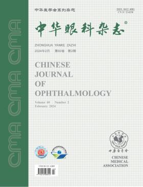
Chinese Journal of Ophthalmology
Volume 60 · Issue 02 · 2024
Chin J Ophthalmol
This issue focuses on the research progress of keratopathy. The responsible editorial committee responsible for the planning, composition and review of this issue is Professor Shi Weiyun.
There are various surgical methods for corneal transplantation, each requiring precise treatment tailored to the characteristics and different layers of corneal opacity or lesions. These layers include the corneal epithelium, stroma (lamellar), and corneal endothelium, with options for full-thickness (penetrating) corneal transplantation or artificial corneal transplantation. However, a current issue in clinical practice is that, regardless of the degree of corneal disease, the availability of corresponding eye bank support, or surgical conditions, classic penetrating (full-thickness) corneal transplantation is universally performed. Alternatively, there is a trend toward adopting technically demanding procedures such as endothelial transplantation and artificial corneal transplantation. This trend has led to increased postoperative complications and the wastage of corneal donor materials. Choosing the appropriate corneal transplant procedure can offer advantages such as preserving more healthy corneal tissue, conserving corneal donors, facilitating rapid vision recovery, and minimizing the risk of immune rejection. Corneologists need to master the indications for various corneal transplant surgeries and systematically perform different corneal transplant procedures based on the surgeon′s skills, hospital conditions, and eye bank conditions. This approach aims to enhance the success rate of the surgery
Endothelial keratoplasty (EK) is an effective therapy for corneal endothelial diseases and mainly includes Descemet stripping endothelial keratoplasty and Descemet membrane endothelial keratoplasty. Compared with penetrating keratoplasty, EK has better vision rehabilitation and a lower rate of allograft rejection after surgery. However, EK poses a series of technical challenges, and varied complications may occur intraoperatively and postoperatively. A successful EK surgery depends on the standardized technical manipulations and the management of surgical complications. In order to standardize the surgical procedure of EK in China, the Cornea Group of Ophthalmology Branch of Chinese Medical Association has made a comprehensive discussion about indications of surgery, preoperative evaluation, surgical manipulations, and the management of complications based on the literature available thus far and clinical practice in China, and eventually established this consensus, which may guide corneal surgeons in performing the EK surgery.
The number of contact lens wearers in China is increasing rapidly. Long-term contact lens wearing may induce or exacerbate tear film instability, and consequently affect the visual quality, ocular surface safety, and compliance of the wearers. To promote the safety and effectiveness of contact lens fitting, and to improve the visual quality and satisfaction of contact lens wearers, the Society of Contact Lens Safety Monitoring & Vision Health of Chinese Health Association has targeted to develop an expert consensus on the diagnosis and treatment of contact lens-associated dry eye. By referring to relevant literature and summarizing clinical experience, the clinical experts in corneal disease, pediatric ophthalmology and optometry have made repeated discussions to form this consensus, including the definition, pathological mechanisms, influencing factors, evaluation, prevention and treatment of contact lens-associated dry eye, for the reference of optometrists and ophthalmologists.
To evaluate the efficacy of 0.05% cyclosporine A eye drops combined with vitamin A palmitate eye gel in the treatment of dry eye associated with meibomian gland dysfunction (MGD).
A single-center, prospective, randomized, parallel controlled trial design was used to include patients diagnosed with MGD-associated dry eye. The patients were randomly divided into three groups and administered with medications binocularly for 12 weeks. The CsA+VA group was given 0.05% cyclosporine A eye drops twice a day and vitamin A palmitate eye gel three times a day. The CsA+HA group was given 0.05% cyclosporine A eye drops twice a day and 0.1% sodium hyaluronate eye drops three times a day. The HA group was given 0.1% sodium hyaluronate eye drops 3 times a day. The OSDI score, tear meniscus height, fluorescein tear break-up time, Schirmer Ⅰ test (without anesthesia), tear film lipid layer thickness, meibomian gland morphology and function examination, and corneal fluorescein sodium staining score were evaluated at baseline, 4, 8, and 12 weeks after the initiation of the treatment, respectively.
A total of 120 patients with MGD-related dry eye met the enrollment criteria, but 10 patients were lost to follow-up; 110 patients were finally included for observation, including 36 patients in the CsA+VA group, 38 in the CsA+HA group and 36 in the HA group. The OSDI score, tear meniscus height, fluorescein tear break-up time and meibomian gland secretion of the 3 groups were significantly improved. At the 12th week of the treatment, the differences of the CsA+VA group [25.45±15.11, (0.30±0.13) mm, (3.72±1.40) s, (5.03±2.52) points] and the CsA+HA group [26.98±16.89, (0.27±0.10) mm, (4.34±1.76) s, (5.11±2.39) points] from the HA group [24.57±11.26, (0.24±0.06) mm, (3.18±1.11) s, (9.11±3.34) points] were statistically significant (P<0.05). Compared with the CsA+HA group [(68.39±26.66) nm], the tear film lipid layer thickness in the CsA+VA group [(72.61±23.65) nm] was significantly increased (P<0.05). In the CsA+VA group, the meibomian gland secretion characters and discharge capacity among patients with severe abnormalities [(6.28±2.59) and (5.89±2.77) points at the 12th week of treatment], moderate abnormalities [(4.27±2.02) and (4.64±2.02) points at the 12th week of treatment] and mild abnormalities [(2.80±0.84) and (2.60±0.55) points at the 12th week of treatment] were significantly different (P<0.05).
0.05% cyclosporine A combined with vitamin A palmitate can significantly improve the symptoms and signs of patients with MGD-related dry eye, especially the tear film lipid layer thickness and the meibomian gland secretion characters and discharge capacity in severe cases.
To investigate the levels of cytomegalovirus (CMV) infection and associated risk factors in corneal transplant recipients who experienced transplant failure.
This was a case-control study. Clinical data from 576 cases (576 eyes) of patients who underwent repeat corneal transplant surgery at the Department of Ophthalmology, Peking University Third Hospital, due to corneal transplant failure from January 2016 to May 2022 were collected. Of these, 305 were male and 271 were female, with a median age of 44.0 (0.7, 91.0) years. The CMV infection rate was analyzed based on the detection of CMV DNA in aqueous humor or corneal tissue during corneal transplant surgery. Patients were divided into the CMV group (CMV DNA positive) and the control group (herpes virus DNA negative). The main research indicators included the CMV infection rate, clinical characteristics, and risk factors in corneal transplant recipients. Chi-square tests and binary logistic analysis were used to compare differences between the two groups in general information, systemic diseases, ocular lesions, ocular surgical history, and local and systemic medications. Odds ratios (OR) and 95% confidence intervals (CI) were calculated for each CMV infection risk factor.
The overall CMV infection rate was 21.9%(126/576), with annual rates ranging from 10.9% to 37.7% from 2016 to 2021. After applying inclusion and exclusion criteria, 378 patients were included in the control trial, with 126 in the CMV group and 252 in the control group. Statistically significant differences between the two groups were observed in systemic immune-related corneal lesions [CMV group: 38 (30.2%), control group: 26 (10.3%)], local immune and inflammatory corneal lesions [CMV group: 46 (36.5%), control group: 40 (15.9%)], congenital corneal opacity [CMV group: 46 (36.5%), control group: 48 (19.0%)] total number of corneal transplants (CMV group: 178 times, control group: 276 times), corneal deep neovascularization crossing the graft [CMV group: 104 (82.5%), control group: 68 (27.0%)] and severe opacity [CMV group: 44 (34.9%), control group: 30 (11.0%)]. Binary logistic regression analysis showed that systemic immune-related corneal lesions (OR=4.044, 95%CI 1.810-9.033, P<0.001), local immune and inflammatory corneal lesions (OR=3.554, 95%CI 1.569-8.052, P=0.002), congenital corneal opacity (OR=2.606, 95%CI 1.216-5.589, P=0.014), total number of corneal transplants (OR=3.206, 95%CI 1.753-5.864, P<0.001), corneal deep neovascularization crossing the graft (OR=8.347, 95%CI 3.967-17.559, P<0.001), and severe opacity (OR=3.063, 95%CI 1.221-7.682, P=0.017) were independent risk factors for CMV infection after corneal transplant.
CMV infection was present in more than 1/5 of corneal transplant recipients who experienced transplant failure. CMV infection after corneal transplant may be related to immune rejection reactions and ocular inflammatory responses. Inflammatory corneal lesions associated with systemic or local immune abnormalities, congenital corneal opacity, and multiple corneal transplants may exacerbate the levels of inflammatory factors during the perioperative period of corneal transplant, increasing the risk of post-transplant CMV infection, leading to the infiltration of deep neovascularization and severe opacity in the cornea.
This study aimed to observe the clinical efficacy of precise suturing of posterior elastic layer fissures guided by intraoperative optical coherence tomography (OCT) in conjunction with anterior chamber puncture and drainage, and corneal thermokeratoplasty for the treatment of severe acute edematous keratoconus.
Non-randomized controlled trial. Data were collected for a study involving 31 cases of acute edematous keratoconus patients who underwent surgical treatment at the Shandong Eye Hospital between June 2017 and July 2021. Among them, there were 30 male and 1 female patients, with an age range of 11 to 32 years and a mean age of (19.80±5.80) years. Eighteen patients in the study group underwent precise suturing of posterior elastic layer fissures guided by intraoperative OCT, in combination with anterior chamber puncture and drainage, and corneal thermokeratoplasty. Thirteen patients in the control group did not undergo suturing. Preoperative visual acuity, corneal edema diameter, corneal thickness, and posterior elastic layer fissure length were collected. Evaluation was performed using slit lamp microscopy, anterior segment OCT, and other methods to assess the time of initial postoperative corneal edema resolution and closure of the posterior elastic layer fissure. Deep lamellar keratoplasty was performed 2 to 4 weeks after edema resolution, and the corneal bed scar repair and visual acuity of the two groups were compared.
In the suturing group, the corneas of all 18 patients were accurately sutured to the deep stromal layer near the posterior elastic layer. The time for corneal edema resolution was 2.50 (1.00, 6.25) days in the suturing group and 7.00 (6.00, 10.50) days in the control group. The fissure healing time was 7.50 (7.00, 12.00) days in the suturing group and 14.00 (9.00, 14.00) days in the control group. The differences were statistically significant (all P<0.05). After 2 weeks, the central corneal thickness decreased to (529.80±174.50) μm in the suturing group and (612.00±205.12) μm in the control group. The suturing group showed accurate corneal suturing to the deep stromal layer near the posterior elastic layer, resulting in central corneal flattening, closure of voids in the stroma, and a significant decrease in corneal thickness. All 18 patients in the suturing group successfully completed deep lamellar keratoplasty, with 6 cases (6/18) experiencing mild graft bed leakage during surgery but without affecting the deep lamellar keratoplasty. One year postoperatively, the visual acuity (logarithm of the minimum resolution angle) was 0.23±0.12 in the suturing group and 0.33±0.11 in the control group, with a statistically significant difference (P<0.05).
In the treatment of severe acute edematous keratoconus, precise suturing of posterior elastic layer fissures guided by intraoperative OCT, in conjunction with anterior chamber puncture and drainage, and corneal thermokeratoplasty, can rapidly alleviate corneal edema and promote the healing of posterior elastic layer fissures. This approach achieves better visual outcomes for subsequent lamellar keratoplasty surgeries. The use of intraoperative OCT guidance allows accurate positioning of the posterior elastic layer fissure in terms of location, direction, and depth of corneal stromal voids, thereby assisting surgeons in precise suturing.
To review the studies related to keratoconus in China, investigate research hotspots and development trends in this field, and provide reference for future research.
This is a bibliometrics study. The relevant literature written in Chinese was retrieved from the WanFang DATA and the China National Knowledge Infrastructure, English articles were collected from the Web of Science Core Collection database. Searched for journal articles related to keratoconus VOSviewer software, CiteSpace, and Bibliometrix in the R language were employed to create the knowledge map. The analysis encompassed the distribution of published journals, research collaboration networks of countries/regions, institutions, and authors. Additionally, core authors, high-frequency keyword co-occurrence, keyword topic maps, and keyword emergence time ranking were examined.
The study ultimately included 1 100 Chinese articles and 668 English articles. Chinese literature and English literature began to increase in 1997 and 2009, respectively, indicating that the field is currently in a developmental stage. The publications involved 244 Chinese journals and 150 English journals, predominantly in the field of ophthalmology. The United States collaborated the most with China, contributing to 123 articles, followed by other countries such as the United Kingdom and Switzerland. Chinese literature and English literature involved 552 and 883 institutions, respectively. The institution with the highest number of Chinese literature publications was the Eye Institute of Shandong First Medical University (63 papers), while Wenzhou Medical University had the highest number of English literature publications (91 papers). Chinese literature involved 2 435 authors, and English literature involved 2 073 authors. The largest collaboration cluster in Chinese literature was formed by the teams of Xie Lixin and Shi Weiyun, while the Gao Hua team formed the largest cluster in English literature. However, collaboration between authors was primarily limited to within each team. A total of 622 and 1 611 keywords were extracted from Chinese and English literature, respectively. The node centrality of the four Chinese keywords, "keratoconus", "cornea", "corneal transplantation" and "myopia" as well as the three English keywords, "keratoconus", "collagen cross-linking" and "penetrating keratoplasty" was greater than 0.1. "Collagen", "riboflavin", "corneal transplantation" and "ultraviolet A" were identified as common core hotspots and important research topics in Chinese and English literature on keratoconus. Keyword emergence analysis indicated that the keywords with the highest intensity of emergence in Chinese and English literature were "myopia" (13.54) and "penetrating keratoplasty" (9.99), respectively. The longest emergence time was observed for "contact lenses" (1995-2006) and "penetrating keratoplasty" (2003-2014).
At present, research on keratoconus in China is on the rise, with research hotspots including pathogenesis, various new surgical methods, and improvement in quality of life. The future research trend mainly focuses on early diagnosis and screening methods, artificial intelligence, biomechanical examination, subclinical keratoconus, and small incision lenticule extraction.
To analyze the early changes in vault height and its influencing factors after implantation of posterior chamber phakic intraocular lenses (pIOL).
A retrospective case series study was conducted, including patients who underwent pIOL implantation at Zhongshan Ophthalmic Center, Sun Yat-sen University, from September 2020 to August 2021, and completed a 3-month follow-up. Data were collected from myopic or myopic astigmatism patients. Preoperative ocular examinations, including Pentacam anterior segment analysis system, Sirius anterior segment analysis system, ultrasound biomicroscopy (UBM), and IOLMaster optical biometry, were performed to measure parameters such as refractive power, corneal curvature, corneal horizontal diameter, anterior chamber volume, anterior chamber depth, pupil diameter, sulcus-to-sulcus diameter (STS), and lens thickness. The degree and position of implanted pIOL, as well as vault height measured by anterior segment optical coherence tomography (AS-OCT) at 1 day, 1 week, 1 month, and 3 months postoperatively, were recorded. Statistical analyses were conducted using repeated measures analysis of variance, Pearson correlation analysis, and multiple linear regression analysis.
A total of 314 patients (314 eyes) were included, with 52 male (16.56%) and 262 female (83.44%) patients, and an average age of (26.44±4.60) years. The preoperative equivalent spherical power was (-8.09±2.41) D. Postoperative vault heights at 1 day, 1 week, 1 month, and 3 months were (671.88±273.02) μm, (652.26±272.21) μm, (615.08±259.69) μm, and (591.14±250.71) μm, respectively, with statistically significant differences among groups (P<0.001). Eyes with vault height>750 μm showed a greater decrease in early postoperative vault height (P<0.001). The eyes implanted with 12.1 mm pIOL had the lowest postoperative vault height, while those with 13.2 mm had the highest (P>0.05). Factors correlated with vault height at 1 day postoperatively included corneal horizontal diameter, anterior chamber depth, preoperative cylinder power, angle degree, lens thickness, and pIOL cylinder power. Factors correlated with vault height at 3 months postoperatively included corneal horizontal diameter, anterior chamber depth, preoperative cylinder power, anterior chamber volume, angle degree, lens thickness, axial length, pIOL spherical and cylinder power. Factors associated with changes in early postoperative vault height included corneal curvature K2, anterior chamber depth, anterior chamber volume, pupil diameter, horizontal STS, vertical STS, axial length, and preoperative spherical power (all P<0.05). Multiple linear regression analysis revealed that lens thickness significantly influenced vault height at 1 day postoperatively, anterior chamber volume significantly influenced vault height at 3 months postoperatively, and pupil diameter significantly influenced changes in early postoperative vault height (all P<0.05).
Vault height after pIOL implantation is unstable in the early postoperative period and gradually decreases within 3 months. A higher baseline vault height is associated with a greater decrease. Anterior chamber volume, pupil diameter, and lens thickness are influencing factors on vault height during the first 3 months postoperatively.
The patient, a 66-year-old male, suffered from redness, blurred vision, photophobia, and tearing in the right eye after being injured by a wooden board. Anti-inflammatory treatment showed poor effectiveness. A 4 mm × 4 mm infiltrate with white deposits on the surface was observed in the central cornea of the right eye. Microscopic examination of corneal scrapings, fungal culture, and in vivo confocal microscopy all indicated fungal infection. The isolated strain was identified as Scedosporium apiospermum through microscopic morphology and confirmed as Petriella setifera by gene sequencing. The patient received corneal debridement combined with routine anti-inflammatory and antifungal treatment in the outpatient clinic. During the follow-up period, the condition continued to improve. Slit lamp examination at the revisit 40 days after the initial diagnosis revealed thinning of the corneal stroma, basic healing of the epithelium, and an increase in uncorrected visual acuity from 0.3 to 0.6.
Different from classical autosomal recessive Wolfram syndrome, Wolfram-like syndrome is an autosomal dominant disorder caused by a heterozygous mutation in the WFS1 gene. In this case, a 7-year-old male child presented to the eye clinic due to vision loss that could not be corrected, discovered during a routine examination. The child had experienced hearing impairment since early childhood, leading to cochlear implantation. Ophthalmic examination revealed optic disc atrophy in both eyes. Optical coherence tomography imaging demonstrated a distinctive thickening of the outer plexiform layer with abnormal layering, characteristic of a single mutation in the WFS1 gene. Subsequent genetic testing identified a de novo heterozygous missense mutation c.2051C>T (p.A684V) in the WFS1 gene, which ultimately led to the diagnosis of Wolfram-like syndrome.
The patient was a 48-year-old male. He went to the Department of Ophthalmology of the First Hospital of Jilin University due to decreased visual acuity in both eyes and tearing for 2 years. A history of systemic illness, ocular trauma, or surgery is denied. Eye examination: the corrected visual acuity of both eyes was 0.6; IOP was 14 mmHg (1 mmHg =0.133 kPa) in the right eye and 15 mmHg in the left eye; The corneal epithelial layer and superficial stromal layer of both eyes are scattered with granular and patchy gray-white opacity (Quintessential Picture 1). In vivo confocal microscopy: flaky highly reflective deposits were seen in the corneal epithelium and shallow stroma of both eyes (Quintessential Picture 2). Clinical diagnosis: Type II granular corneal dystrophy in both eyes.
Corneal diseases are prevalent eye conditions in China, and the lack of effective treatment in the short term can lead to blindness. However, delivering drugs to the cornea safely and effectively poses a significant challenge due to the presence of ocular barriers and clearance mechanisms. Conventional drug delivery methods exhibit low bioavailability, making it difficult to achieve therapeutic effects. Microneedles, with their ability to penetrate ocular surface barriers effectively, offer a low-invasive and highly promising drug delivery technology. This article introduces the main delivery barriers on the ocular surface, classifies microneedles, and highlights the latest developments in the treatment of corneal diseases. Finally, the potential challenges of applying microneedle delivery systems to the ocular surface are analyzed, aiming to provide insights for the clinical application of microneedles in corneal diseases.
Myopia is a global public health issue, particularly prevalent in China, with a rising trend in recent years. The increased use of computers, smartphones, and video display terminals has led to frequent dry eye symptoms, such as blinking, among myopic students. Studies have revealed a higher incidence of dry eye in myopic children compared to emmetropic children, significantly impacting their learning and quality of life. However, ophthalmologists have traditionally focused more on the prevention and control of myopia, often neglecting ocular surface health awareness in children. It is essential to understand the potential impact of myopia on dry eyes in children and whether there is a difference in dry eye prevalence. This article reviews the current state of research on childhood myopia-related dry eye, encompassing epidemiology, pathogenesis, and risk factors, aiming to provide clinical reference for intervention, prevention, and precise treatment of dry eyes in myopic children.
Femtosecond laser-assisted arcuate keratotomy (FSAK) is an effective, long-term stable, and safe surgical procedure for astigmatism correction. Its application in cataract surgery and the correction of astigmatism combined with cataracts is becoming increasingly widespread. Apart from its application in key steps of cataract surgery, both domestic and international research indicates that the introduction of femtosecond laser technology has enhanced the precision of arcuate incisions in keratotomy, reducing the risk of intraoperative or postoperative complications. This has significantly contributed to correcting low to moderate astigmatism in patients undergoing cataract surgery. However, the effectiveness of FSAK relies on obtaining more accurate incision parameters and refining surgical planning. This article provides a comprehensive review of the corrective effects, limitations, and preoperative planning aspects of femtosecond laser-assisted arcuate keratotomy.
Posterior corneal elastic layer detachment (DMD) is a pathological state in which the posterior corneal elastic layer is separated from the corneal stromal layer, which is manifested by corneal edema and opacity in the separated area. It is one of the serious complications of endocular surgery, and in severe cases, corneal endothelial decompensation may occur. Based on the anatomical structure and physiological function of the posterior corneal elastic layer, this paper systematically explains the causes and risk factors, clinical manifestations, diagnosis and classification, treatment principles and methods of DMD, in order to provide reference and help for ophthalmologists to master the rapid diagnosis methods and effective treatment techniques of DMD, and improve the clinical diagnosis and treatment level.
CURRENT ISSUE