Chinese Neurosurgical Journal
Volume 06 · Issue 03 · 2020
Chin Neurosurg J
- Sections
- Research
- Review
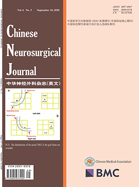
When utilizing the retrosigmoid approach (RA), accurately identifying the transverse and sigmoid sinus transition (TSST) is a key procedure for neurosurgeons, especially in developing countries restricted by the lack of expensive devices, such as the neural navigation system and the three-dimensional volumetric image-rendered system. Before operations, a computed tomography scan is a common and cost-effective method of checking patients who suffer lesions located at the cerebellopontine angle. Therefore, we present a technique using only high-resolution computed tomography to identify the transverse and sigmoid sinus transition.
This retrospective study included 35 patients who underwent retrosigmoid approach operations to resect an acoustic neurinoma with the assistance of our technique. In brief, our technique contains 4 steps: (1) All patients’ 1-mm, consecutive, high-resolution computed tomographic images that clearly displayed landmarks, such as the inion, lambdoid suture, occipitomastoid suture, and the mastoid emissary foramen, were investigated initially. (2) We selected two particular slices (A and B) among all of these high-resolution computed tomographic images in which scanning planes were parallel with the line drawn from the root of the zygoma to the inion (LZI). Slice A contained both the root of the zygoma and the inion simultaneously, and slice B displayed the mastoid emissary foramen. (3) Four points (α, β, γ, δ) were arranged on slices A and B, and point α was located at the inner surface of the skull, which represents the posterior part of the sulci of the sigmoid sinus. Point β was located at the outer surface of the skull, and the line connecting them was perpendicular to the bone. Similarly, on slice B, we labeled point γ as the point that represents the posterior part of the sulci of the sigmoid sinus at the inner surface and point δ as the point located at the outer surface of the skull, and the line connecting them was also perpendicular to the bone. The distances between point β and the lambdoid suture/occipitomastoid suture and between point δ and the mastoid emissary foramen were calculated for slices A and B, respectively. (4) During the operation, a line indicating the LZI was drawn on the bone with ink when the superficial soft tissue was pushed away, and this line would cross the lambdoid suture/occipitomastoid suture. With both the crosspoint and the distance obtained from the high-resolution CT images, we could locate point β. We also used the same method to locate point δ after revealing the mastoid emissary foramen. The line connecting point β and point δ indicated the posterior border of the sigmoid sinus, and the intersection between the line and LZI indicated the inferior knee of the transverse and sigmoid sinus transition (TSST).
All 35 patients underwent the RA craniectomies that were safely assisted by our technique, and neither the sigmoid sinus nor the transverse sinus was lacerated during the operations.
Our cost-effective technique is reliable and convenient for identifying the transverse and sigmoid sinus transition (TSST) which could be widely performed to guarantee the safety of RA craniectomy.
Endonasal endoscopic skull base surgery has undergone rapid technological developments and is now widely performed, but its strengths and weaknesses deserve further investigation and deliberation. This study was performed to investigate the surgical indications, complications, and technical advantages and disadvantages of endonasal endoscopic skull base surgery.
The clinical data of 1886 endoscopic endonasal skull base surgeries performed in our ward at Beijing Tiantan Hospital from June 2006 to June 2016 were retrospectively analyzed.
One thousand ninety-three (73.4%, 1490) pituitary adenomas, 54 (24.9%, 217) chordomas, 28 (80.0%, 35) craniopharyngiomas, and 15 (83.3%, 18) meningiomas underwent total resection. Two patients died postoperatively, both having pituitary adenomas. Other postoperative complications included olfactory disorders (n = 226, 11.9%), postoperative cerebrospinal fluid leakage (n = 78, 4.1%), hypopituitarism (n = 74, 3.9%), diabetes insipidus (n = 64, 3.4%), intracranial infection (n = 36, 1.9%), epistaxis (n = 24, 1.3%), vascular injury (n = 8, 0.4%), optic nerve injury (n = 8, 0.4%), and oculomotor movement impairment (n = 4, 0.2%). In total, 1517 (80.4%) patients were followed up for 6 to 126 months (average, 42.5 months) postoperatively. A total of 196 (13.2%) pituitary adenomas and 13 (37.1%) craniopharyngiomas recurred but no meningiomas recurred. Chordomas recurred in 97 (44.7%) patients, in whom 5-year survival rate was 65%.
Endoscopic surgery is an innovative surgical technique and the first choice for most midline extradural lesions such as chordomas, and an excellent choice for pituitary adenomas. It probably will be a good technique for many kinds of craniopharyngiomas and a common technique for most of skull base meningiomas, so the surgical indications of these cases should be chosen carefully to make good use of its respective advantages.
Deregulation of miRNA-21 expression has been reported to be associated with vascular smooth muscle behavior and cytoskeletal stability. This study is aimed to investigate the density of serum miRNA-21 in patients with different phases of intracranial aneurysms (IAs) and explore its warning function for IA rupture.
A total of 16 in 200 IA patients were selected and categorized into 4 groups based on the phase of IA. Microarray study was carried out using serum miRNA and differentially expressed miRNAs were identified. Another 24 samples from a cohort of 360 patients were added and real-time polymerase chain reaction (RT-PCR) was performed on expanded sample size (n = 40) for miRNA-21 validation. Potential gene targets of miRNA-21 were screened out from Gene Ontology (GO) database and literatures.
Microarray study identified 77 miRNAs with significantly different expression levels between experimental groups and the control group. RT-PCR assays validated significant downregulation of miRNA-21 in experimental groups, among which miRNA-21 expression level of daughter aneurysm group decreased the most. Bioinformatic analyses revealed that several target genes related with miRNA-21 may be involved in IA formation and rupture.
This study suggested that miRNA-21 had a protective effect for intracranial vascular wall against remodeling and warning function for intracranial aneurysm rupture. Significant suppression of serum miRNA-21 in IA patients may provide diagnostic clues for aneurysm rupture and guide clinical intervention.
Recent advances in surgical and neuroprotective strategies could effectively manage the pathophysiological progression of subarachnoid hemorrhage (SAH). However, pulmonary dysfunction frequently occurs in SAH patients with an increased risk of unsatisfactory outcomes. Based on the similar microvascular structures in the blood-air barrier and blood-brain barrier and possible brain-lung crosstalks, we believe that pericytes may be involved in both neurological and pulmonary dysfunction after SAH.
In our experiments, platelet-derived growth factor B (PDGF-B) retention motif knockout (PDGF-Bret/ret) mice and adeno-associated virus PDGF-B were employed to show the involvement of pericyte deficiency and PDGF-B expression. Neurological score, SAH grade, hematoxylin-eosin staining, and PaO2/FiO2 ratio analysis were performed to evaluate the neurological deficits and pulmonary functions in endovascular perforation SAH models at 24 h after surgery, as well as western blotting and immunofluorescence staining for underlying molecular expressions.
We found that neonatal PDGF-Bret/ret mice exhibited pulmonary atelectasis 12 h after birth. Further investigation showed a decrease in PaO2/FiO2 and lung-specific surfactant proteins in adult PDGF-Bret/ret mice. These dysfunctions were much worse than those in wild-type mice at 24 h after SAH. PDGF-B overexpression alleviated pulmonary dysfunction after SAH.
These results suggested pulmonary dysfunction after SAH and the pivotal role of PDGF-B signaling for the pathophysiological process and future therapeutic targets of pulmonary injury treatment after SAH. Further studies are needed for pathophysiological investigations and translational studies on pulmonary injuries after SAH.
Our study aimed to determine the effect of the neutrophil-lymphocyte ratio on the prognosis of adult patients with acute stroke. We searched the Web of Science, PubMed, Embase, Cochrane Library, and China National Knowledge Infrastructure databases and selected all of the potentially eligible studies. From the included studies, we extracted characteristics such as the stroke type and acquisition time until routine blood collection and the odds ratios across studies. The 95% confidence intervals and odds ratios were pooled to calculate the effect size for the neutrophil-lymphocyte ratio in acute stroke patients. We defined poor function outcomes according to the modified Rankin Scale ≥ 3 or Glasgow Outcome Scale< 3.Thirteen studies with 4443 patients were included in our analysis, including 7 ischemic and 6 hemorrhagic stroke studies. The pooled odds ratios for poor functional outcome at 3 months with a higher neutrophil-lymphocyte ratio in acute ischemic and hemorrhagic patients were 1.689 (95% CI = 1.184-2.409, p < 0.001) and 1.125 (95% CI = 1.022-1.239, p < 0.001), respectively, and the overall pooled odds ratio for poor functional outcome following stroke was 1.257 (95% CI = 1.146-1.379, p < 0.001). At the same time, the overall combined odds ratio for death at 3 months was 1.632 (95% CI = 1.155-2.306, p < 0.001).The neutrophil-lymphocyte ratio, an easily calculated marker, plays a predictive role in the short-term outcomes of adult patients (mean age ≥ 50 years) following acute ischemic and hemorrhagic stroke.
To conduct a systematic review and meta-analysis and evaluate the effect of tranexamic acid in patients with traumatic brain injury. PubMed, EMBASE, and CENTRAL (Cochrane Central Register of Controlled Trials) were searched to identify randomized controlled trials and evaluate the effect of tranexamic acid in traumatic brain injury patients. The primary outcome was mortality. Two reviewers extracted the data independently. The random effect meta-analysis was used to estimate the aggregate effect size of 95% confidence intervals. Six randomized controlled trials investigating tranexamic acid versus placebo and 30073 patients were included. Compared with placebo, tranexamic acid decreased the mortality (RR = 0.92; 95% CI, 0.87-0.96; p < 0.001) and growth of hemorrhagic mass (RR = 0.78; 95% CI, 0.61-0.99; p = 0.04). However, tranexamic acid could not decrease disability or independent, neurosurgery, vascular embolism, and stroke. Current evidence suggested that compared with placebo, tranexamic acid could reduce mortality and growth of hemorrhagic mass. This finding indicated that patients with traumatic brain injury should be treated with tranexamic acid.
Globally, the discipline of neurosurgery has evolved remarkably fast. Despite being one of the latest medical specialties, which appeared only around hundred years ago, it has witnessed innovations in the aspects of diagnostics methods, macro and micro surgical techniques, and treatment modalities. Unfortunately, this development is not evenly distributed between developed and developing countries. The same is the case with neurosurgical education and training, which developed from only traditional apprentice programs in the past to more structured, competence-based programs with various teaching methods being utilized, in recent times. A similar gap can be observed between developed and developing counties when it comes to neurosurgical education. Fortunately, most of the scholars working in this field do understand the coherent relationship between neurosurgical education and neurosurgical practice. In context to this understanding, a symposium was organized during the World Federation of Neurological Surgeons (WFNS) Special World Congress Beijing 2019. This symposium was the brain child of Prof. Yoko Kato—one of the eminent leaders in neurosurgery and an inspiration for female neurosurgeons. Invited speakers from different continents presented the stages of development of neurosurgical education in their respective countries. This paper summarizes the outcome of these presentations, with particular emphasis on and the challenges faced by developing countries in terms of neurosurgical education and strategies to cope with these challenges.
CURRENT ISSUE