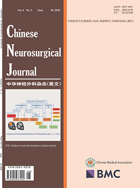
Chinese Neurosurgical Journal
Volume 06 · Issue 02 · 2020
Chin Neurosurg J
- Sections
- Research
- Review
- Case Report
The prevalence of low back pain with radiculopathy in general population varies from 9.9% to 25%, which can be due to bony narrowing of the lateral recess or due to prolapsed intervertebral disc. Transforaminal epidural injection of a mixture of long-acting anaesthetic (bupivacaine) and particulate steroids (depomedrol) has been a treatment modality in patients not responding to initial physiotherapy and neuropathic pain medications.
To analyze the effectiveness of transforaminal epidural steroid injection (TFESI) in the treatment of low back pain with radiculopathy, a retrospective case series evaluating the records of patients that received TFESI (1 mL 0.5% bupivacaine +1 ml/40 mg depomedrol) under C-arm guidance from January 2015 to December 2018 (3 years) at Upendra Devkota Memorial-National Institute of Neurological and Allied Sciences (UDM-NINAS), their lumbosacral MRI and the pre-procedure, 1-week and 3-month numeric pain rating scale, were analyzed. Successful treatment (reduction of pain scale by more than 50% of baseline at 3 months) in the patients with bony recess stenosis and those with prolapsed intervertebral disc was compared.
Out of 67 patients that received TFESI, 35 (52.23%) could be followed up. The mean age was 55.8 ± 14.39 years and 51.3% were females. 68.57% had L5 and 20% had S1 radiculopathy. Bony recess stenosis was seen in the aged 40% and PIVD was the cause of radiculopathy in 60%. The median duration of radicular pain prior to intervention was 3 months. TFESI was effective as the mean numeric pain scale before injection was 8.97 ± 1.32 which reduced to 3.91 ± 3.23 (paired t test p value < 0.001) at 1 week post injection and 3.23 ± 3.34 (paired t test p value < 0.001) at 3 months post injection. Twenty-six of the 35 patients (75.29%) had more than 50% pain relief compared to baseline at 3 months and were satisfied. Nine patients continued to have pain; however, only one required a surgical intervention. The effectiveness of TFESI was not significantly different in different ages (Fisher’s exact test p value 0.182) and in different anatomic levels (Fisher’s exact test p value 0.241). Six out of eight patients with bony recess stenosis benefited as compared to 14 out of 19 patients with PIVD, though it was not statistically significant (Fischer’s exact test p value 0.688). There were no adverse events recorded.
TFESI is a safe and efficacious treatment modality in patients with radicular low back pain especially in aged patients in whom surgery under general anaesthesia is not free from risk.
The purpose of this study was to use the modified Delphi method to identify the influencing factors of health-related quality of life (HRQoL) in patients with unruptured intracranial aneurysms (UIAs) after endovascular treatment.
A modified Delphi method to obtain expert consensus on the content of potential influencing factors of HRQoL in patients with UIAs treated by endovascular intervention was employed. The research team consists of three neuroradiologists and one epidemiologist from Xuanwu Hospital of Capital Medical University. They randomly selected 21 well-known experts in cerebrovascular disease diagnosis and treatment as participating experts. The importance of the indicator is based on the 5-Likert scale. The standard deviation (SD), coefficient of variation (CV), mean ( ), and minimum and maximum scores of each indicator were calculated. The consistency was described by Kendall coefficient of concordance with a p value < 0.05 indicating that the expert consistency was high.
Twenty-one and 18 questionnaires were responded in 2 rounds, with effective response rates of 85.7% and 100.0%, respectively. The average authoritative coefficient (Cr) of all 21 experts was 0.88, familiarity with the indicators (Cs) was 0.82, and the judgment basis of the indicators (Ca) was 0.94. Eventually, the  values of arterial puncture hematoma, hyperlipidemia, gender, marital status, and hospitalization for other diseases were lower than 3.5; CV for marital status and gender was higher than 0.35. The Kendall coefficient of concordance in the first round was 0.19 (p < 0.001), and the second round was 0.15 (p < 0.001).
In this study, the factors affecting the recovery of HRQoL after endovascular treatment in patients with UIAs were analyzed by the modified Delphi method, which provided a valuable evidence for the clinical management and daily life guidance for UIAs patients.
Piriformis syndrome accounts for approximately 6% of patients who present with sciatic pain. There are many treatment options ranging from physical therapy, to trigger point injections, to surgical intervention. We discuss a surgical method that represents a minimally invasive technique for the treatment of piriformis syndrome.
We describe a novel operative approach and technique for release of the piriformis muscle in the treatment of piriformis syndrome. Described are the preoperative planning, incision and approach, and technique for identifying and releasing the piriformis muscle.
Three patients were treated for piriformis syndrome using the described technique. Each patient displayed successful relief of their symptoms immediately following the surgical procedure and at delayed follow-up.
Early experience with our method of piriformis release suggests that it is well suited for the treatment of piriformis syndrome. The novel integration of pre-operative trigger point localization coupled with intraoperative neuromonitoring allows effective pain relief with minimal morbidity.
Gliomas are the most common primary malignant brain tumors and have a poor prognosis. Early detection of gliomas is crucial to improve patient outcomes. Urine accumulates systematic body changes and thus serves as an excellent early biomarker source.
At the biomarker discovery phase, we performed a self-controlled proteomics analysis by comparing urine samples collected from five glioma patients at the time of tumor diagnosis and after surgical removal of the tumor. At the biomarker validation phase, we further validated some promising proteins using parallel reaction monitoring (PRM)-based targeted proteomics in another cohort, including glioma, meningioma, and moyamoya disease patients as well as healthy controls.
Using label-free proteome quantitation (LFQ), we identified twenty-seven urinary proteins that were significantly changed after tumor resection, many of which have been previously associated with gliomas. The functions of these proteins were significantly enriched in the autophagy and angiogenesis, which are associated with glioma development. After targeted proteomics validation, we identified a biomarker panel (AACT, TSP4, MDHM, CALR, LEG1, and AHSG) with an area under the curve (AUC) value of 0.958 for the detection of gliomas. Interestingly, AACT, LEG1, and AHSG are also potential cerebrospinal fluid or blood biomarkers of gliomas.
Using LFQ and PRM proteome quantification, we identified candidate urinary protein biomarkers with the potential to detect gliomas. This study will also provide clues for future biomarker studies involving brain diseases.
The prevalence rates of freezing of gait (FOG) in Parkinson’s disease (PD) vary widely, ranging from 14.0 to 55.1%. Our aim is to calculate the overall prevalence of FOG in all PD patients with different disease durations and severities.
Using Medline/PubMed/Embase, we carried out a systematic literature search for studies reporting the PD and clinically relevant FOG.
After primary screening, a total of 35 studies were identified and further analyzed for inclusion into the analysis, and 29 studies fulfilled the quality criteria and included in this meta-analysis. The overall prevalence of FOG in PD was 39.9% (95% CI 35.3-44.5%). The FOG identified by the freezing of gait questionnaire item 3 may be more prevalent (43.8%, 95% CI 38.5-49.1%) than the FOG identified by the Unified Parkinson’s Disease Rating Scale item 14 (36.0%, 95% CI 29.0-43.1%). Disease duration and severity are both the clinical features associated with the FOG. The highest FOG prevalence rate in PD patients was seen in patients with disease durations ≥ 10 years, at 70.8%, followed that of PD patients with disease durations ≥ 5 years (53.3%), and PD patients with disease durations < 5 years (22.4%). FOG presented in 28.4% of PD patients with Hoehn and Yahr staging (H&Y) score ≤ 2.5, and in 68.4% of PD patients with H&Y score ≥ 2.5.
This meta-analysis confirms that the prevalence of FOG in PD is considerable, and highlights the need for accurate identification of FOG in PD.
New discoveries based on genetic and epigenetic evidence have significantly expanded the understanding of diffuse gliomas. Molecular biomarkers detected in diffuse gliomas are not only potential targets for radiotherapy, chemotherapy, and immunotherapy, but are also able to guide surgical treatment. Previous studies have suggested that the optimal extent of resection of diffuse gliomas varies according to the expression of specific molecular biomarkers. However, the specific guiding role of these biomarkers in the resection of diffuse gliomas has not been systemically analyzed. This review summarizes several critical molecular biomarkers of tumorigenesis and progression in diffuse gliomas and discusses different strategies of tumor resection in the context of varying genetic expression. With ongoing study and advances in technology, molecular biomarkers will play a more important role in glioma resection and maximize the survival benefit from surgery for diffuse gliomas.
Endolymphatic sac tumor (ELST) is one of neuroectodermal tumor which arising from endolymphatic sac and duct. It is actually quite rare, with less than 200 cases reported. Although ELST presents benign appearance in histopathology, it can present aggressive destructive behavior in clinical. The cornerstone of treatment for ELST is complete surgical excision. However, it is almost impossible to completely resect the advanced stage tumor. There is still controversy about other treatments, such as radiotherapy and gamma knife surgery.
A 47-year-old man was admitted in The First Affiliated Hospital of Fujian Medical University with a 7-year history of progressive hearing loss and near 6-month repeated attacks of headache. Preoperative CT revealed a massive intracranial lesion and associated hydrocephalus. MR scanning demonstrated a 7.2 cm × 4.6 cm × 4.2 cm bulky mass located in left-sided posterior cranial fossa and temporo-occipital region which showed hyperintensity on T1-weighted images and mixed signal intensity on T2-weighted images. There was no neither clinical manifestation nor family history of Von Hippel-Lindau syndrome (VHL).Due to the mass that was large and invading the bone of skull base, it was difficult to extirpate surgically, so the ventriculoperitoneal shunt combined with local biopsy was performed. The postoperative pathology and immunohistochemical findings confirmed the lesion was an endolymphatic sac tumor. After operation, the patient regularly received radiotherapy.
The widely accepted management of ELST is complete surgical resection. However, it is difficult for surgeons to achieve radical resection with late-stage ELST. Currently, there is much dispute about the role of radiotherapy for the management of ELST in academic circles. In this case where the mass cannot be surgical removed, radiotherapy has the curative effect for ELST in terms of disease control and quality of life.
Invasive sphenoid sinus aspergillosis is a rare but life-threatening condition usually found in immunocompromised patients. When involving cavernous sinus and surrounding structures, patients are frequently misdiagnosed with a neoplasm or sellar abscess. Timely diagnosis and intervention are crucial to patients’ outcomes. The objective of this study is to review cases of invasive sphenoid sinus aspergillosis to describe disease manifestations, imaging features, treatment, and outcome.
We describe four patients with invasive sphenoid sinus aspergillosis misdiagnosed as sellar tumors preoperatively. The mass was completely removed in three patients and partially removed in one patient microscopically. Pathological examinations confirmed Aspergillus in all cases. All four patients received anti-fungal agents postoperatively. There was no recurrence at the time of each patient’s follow-up date. One patient with complete resection was lost to follow-up while the other three patients’ neurologic function improved. Additionally, we performed a systematic review regarding invasive sphenoid sinus aspergillosis of existing English literature.
With regard to clinical symptoms, headache, vision impairment, and ophthalmoplegia were observed in over half of the patients in the literature. A sellar mass with bone destruction on CT and involvement of cavernous sinus is highly suggestive of invasive fungal sphenoid sinusitis. Immediate surgical removal of the lesion is recommended for invasive sphenoid sinus aspergillosis to preserve nerve function and increase the likelihood of survival.
CURRENT ISSUE