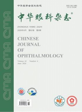
Chinese Journal of Ophthalmology
Volume 62 · Issue 06 · 2026
Chin J Ophthalmol
This issue focuses on the research progress of low vision. The responsible editorial committee responsible for the planning, composition and review of this issue is Professor Lu Fan.
Low vision rehabilitation is an indispensable and integral part of the national eye health initiative, and it is directly related to the quality of life of tens of millions of people with visual impairment in China. Currently, the global prevalence of low vision continues to rise, and China has a large patient base of low vision. However, the accessibility of rehabilitation services remains severely inadequate, and the vast majority of people with visual disability cannot receive standardized visual rehabilition services. Based on the development history and current challenges of low vision rehabilitation globally and in China, this editorial clearly points out that clinical medical institutions are the first gateway of the low vision rehabilitation system, emphasizes the core role of ophthalmologists in the referral system and the superior efficacy and value of multidisciplinary team care, and proposes a patient-centered rehabilitation paradigm integrating science and humanity based on functional visual assessment. Meanwhile, it puts forward specific suggestions on strengthening grassroots referral coordination, accelerating the training of specialized professionals, and promoting the translation and application of new technologies such as artificial intelligence, aiming to provide practical guidance for the high-quality development of low vision rehabilitation in China.
Child and adolescent myopia has emerged as a prominent public health issue in China, characterized by trends of earlier onset and increasing severity. The prevention and control of myopia in children and adolescents present substantial challenges to public health. Driven by robust national policies, several distinctive technical guidance documents have been issued in recent years within relevant professional domains. Nevertheless, notable gaps persist in the quantification of evidence quality and intervention efficacy, multidimensional combination strategies, and the provision of specific operational guidance for clinical and public health practice. In response, the Visual Health (Myopia Prevention and Control) Study Group of the Child and Adolescent Health Branch of the Chinese Preventive Medicine Association convened a multidisciplinary panel comprising clinical experts, public health specialists, and methodologists engaged in clinical practice guideline development. Adhering rigorously to international standards for guideline development, the panel formulated eight clinical questions spanning three core dimensions, including environmental regulation, behavioral modification, and pharmacological/optical correction, and subsequently developed eight corresponding evidence-based recommendations. These recommendations aim to provide more precise and systematic evidence-based guidance for myopia prevention and control in clinical and public health practice, thereby enhancing the scientific validity and effectiveness of myopia management efforts in Chinese children and adolescents.
To investigate the prevalence and etiologies of blindness and visual impairment in China from 1990 to 2021, and to provide references for formulating prevention and treatment strategies for blindness and visual impairment.
This study was an ecological trend study based on the Global Burden of Disease (GBD) 2021 database. Data on the number of cases, prevalence, age-standardized prevalence and etiologies of blindness and visual impairment in China from 1990 to 2021 were extracted. The prevalence and changes in etiological composition in different genders and age groups were analyzed. Joinpoint regression model was used to calculate the Average Annual Percentage Change (AAPC). Decomposition analysis was performed to quantify the contributions of population aging, population growth and epidemiological change to the changes in the number of cases.
In 2021, the number of cases of blindness, severe visual impairment and moderate visual impairment in China was 8.768 2 million, 4.178 9 million and 47.757 2 million, respectively, which increased by 64.90%, 129.47% and 139.80% compared with 1990, with AAPCs of 1.70%, 2.87% and 2.89%, respectively (all P<0.05). The age-standardized prevalence rates were 465.14 per 100 000 population, 214.09 per 100 000 population and 2 501.72 per 100 000 population, respectively, which showed changes of -28.63%, -8.73% and+7.74% compared with 1990, with AAPCs of -1.03% (P<0.05), -0.15% (P=0.281) and 0.28% (P<0.05), respectively. The prevalence of visual impairment was higher in females than in males. In 2021, the highest number of cases of blindness and severe visual impairment was in the 70-74-year age group; the highest number of cases of moderate visual impairment was in the 65-69-year age group. Population aging contributed 133.40%, 87.49% and 68.54% to the changes in the number of cases of blindness, severe visual impairment and moderate visual impairment, respectively; population growth contributed 39.82%, 23.78% and 22.18%, respectively; epidemiological change contributed -73.22%, -10.56% and 9.28%, respectively. Refractive error and cataract were the top two etiologies of blindness and visual impairment.
The number of cases of blindness and visual impairment in China showed an upward trend from 1990 to 2021. With population growth and aging, the disease burden will further increase. Focusing on cataract in the elderly and refractive error in children and adolescents is an important strategy to reduce the burden of blindness and visual impairment in China.
To assess the prevalence of myopia and its main influencing factors among Chinese primary and secondary school students using meta-analysis.
A meta-analysis was conducted. Databases including CNKI, Wanfang Data, China Biology Medicine Database, PubMed and Web of Science Core Collection were electronically searched to collect cross-sectional studies on influencing factors of myopia in Chinese primary and secondary school students published from January 2013 to October 2024. Two researchers independently screened the literature, extracted data and assessed the methodological quality based on the Agency for Healthcare Research and Quality (AHRQ) criteria. Meta-analysis was performed using Stata 18.0 software to calculate the pooled prevalence and the odds ratios (OR) with 95% confidence intervals (CI) for each influencing factor. Heterogeneity test, publication bias assessment and sensitivity analysis were also conducted.
A total of 76 studies were included, covering 31 provinces, autonomous regions and municipalities directly under the Central Government of China, with a cumulative sample size of 1, 823, 018 participants. Eight main influencing factors of myopia were analyzed. The meta-analysis results showed that the risk of myopia in female primary and secondary school students was 1.45 times that in male students (OR=1.45, 95%CI:1.29-1.63), and the risk in secondary school students was 4.11 times that in primary school students (OR=4.11, 95%CI:3.76-4.50). Parental history of myopia (at least one parent affected), excessive daily use of electronic devices, and incorrect eye use posture were identified as risk factors (P<0.05). In contrast, performing eye exercises ≥10 times per week, engaging in outdoor activities ≥ 5 hours per week, and sleeping > 8 hours per day were protective factors (P<0.05). Sensitivity analysis indicated that the results were robust.
The prevalence of myopia among Chinese primary and secondary school students remains high, and the spectrum of influencing factors exhibits distinct "Chinese context" characteristics: the harm of high-intensity near-work driven by academic pressure far outweighs that of isolated electronic screen exposure, while poor reading and writing postures, specifically deviations from the "One Fist, One Chi, One Cun" standardized posture guidelines, significantly elevate myopia risk. Given that the development of myopia is jointly influenced by genetic, behavioral, and environmental factors, it is recommended that while drawing upon international experiences, we fully leverage the strengths of indigenous institutional advantages, such as Chinese eye exercises, and embed standardized posture education into daily classroom routines. Ultimately, the goal is to construct a "society-school-family" trinity prevention and control system with Chinese characteristics, actively guide primary and secondary school students in cultivating healthy eye-use habits, and further strengthen the implementation intensity of comprehensive myopia intervention measures.
To analyze the genetic and clinical characteristics of patients with PAX6-associated ocular diseases (PAOD).
This retrospective case series study recruited 40 PAOD patients from 26 families at Peking Union Medical College Hospital between January 2015 and March 2025. Medical history and ophthalmic examination results were collected, including visual acuity, color fundus photography, fundus autofluorescence, optical coherence tomography (OCT), and B-scan ultrasonography. Peripheral blood samples were obtained from patients and their family members for DNA extraction. The PAX6 pathogenic variants were detected using next-generation sequencing (NGS), multiplex ligation-dependent probe amplification (MLPA), and quantitative real-time quantitative PCR (qPCR), followed by segregation analysis in pedigrees.
A total of 40 patients from 26 unrelated families were identified, with an equal distribution of 20 males and 20 females. The age at first visit ranged from 0.5 to 59.0 years (median: 25.5 years). Best corrected visual acuity ranged from no light perception to 0.4 (median: 0.1). Among the cases, 23 patients were diagnosed with congenital aniridia, while 17 patients had non-aniridia phenotypes, including anterior segment dysgenesis, Peters anomaly, and foveal hypoplasia. Foveal hypoplasia was a common clinical manifestation (100%, 23/23) in PAOD patients, with other common manifestations including nystagmus (94.9%, 37/39), cataract (80.0%, 28/35), and glaucoma (59.3%, 16/27). Twenty-five PAX6 pathogenic variants were identified, among which 9 novel variants were reported in this study, including 3 missense mutations, 3 large chromosomal rearrangements, 1 frameshift mutation, 1 splicing variant, and 1 small deletion. Truncating mutations (68.7%) were the most common variant type in aniridia patients, whereas missense mutations (70.0%) predominated in other PAX6-associated ocular diseases, showing a significant difference (P<0.001).
PAOD exhibits high phenotypic heterogeneity, with foveal hypoplasia as a common feature and congenital aniridia being the most prevalent phenotype. Nystagmus combined with multiple ocular structural abnormalities are key clinical features suggestive of PAOD. This study expands the clinical and genetic spectrum of PAOD and provides further insights into genotype-phenotype correlations.
To compare the clinical efficacy and safety of phacoemulsification with goniosynechialysis (Phaco-GSL) versus Phaco-GSL combined with excisional goniotomy using the Kahook dual blade (KDB) in the treatment of primary angle-closure glaucoma with cataract (PACG-C).
A prospective non-randomized case-control study was conducted. Patients with PACG-C scheduled for combined cataract extraction and anti-glaucoma surgery at the Eye Hospital of Shandong First Medical University from March 2023 to December 2024 were enrolled. Based on the disease severity, they were assigned to either the Phaco-GSL-KDB group or the Phaco-GSL group for the corresponding surgical procedure. The visual acuity, intraocular pressure (IOP), number of anti-glaucoma medications used, visual field, and surgical success rate were recorded and compared between the two groups preoperatively, at 1 day, 1 week, 1 month, 3 months, 6 months, and 12 months postoperatively. The paired t-test, Wilcoxon signed-rank test, independent samples t-test, Mann-Whitney U test, Chi-square test, and Friedman test were employed for statistical analysis.
A total of 101 patients (116 eyes) with PACG-C who met the inclusion criteria were enrolled, including 32 males (34.81%) and 69 females (65.19%), with a mean age of (67.28±7.07) years. There were 56 patients (61 eyes) in the Phaco-GSL-KDB group and 45 patients (55 eyes) in the Phaco-GSL group. In the Phaco-GSL-KDB group, the number of preoperative anti-glaucoma medications was higher than that in the Phaco-GSL group [3.00 (2.00, 3.00) vs. 2.00 (1.00, 3.00)], while the mean deviation was lower [-23.68 (-28.90, -12.81) dB vs.-12.96 (-23.18, -5.27) dB], with differences being statistically significant (both P<0.05). There were no statistically significant differences between the two groups in baseline parameters such as age, visual acuity, and IOP (all P>0.05). At the 12-month follow-up, the visual acuity improved in both groups compared to preoperative levels (both P<0.05). The IOP decreased in both groups compared to preoperative levels, and the Phaco-GSL-KDB group maintained lower IOP than the Phaco-GSL group at all follow-up time points. At 12 months postoperatively, the reduction in IOP was 47.60% in the Phaco-GSL-KDB group vs. 38.01% in the Phaco-GSL group (both P<0.05). The mean deviation improved, and the pattern standard deviation (PSD) decreased in both groups (all P<0.05). The reduction in PSD in the Phaco-GSL-KDB group was 2.8%. Both the complete success rate and conditional success rate were higher in the Phaco-GSL-KDB group compared to the Phaco-GSL group (both P<0.05). Specifically, the conditional success rate [defined as IOP≤18 mmHg (1 mmHg=0.133 kPa) at 12 months postoperatively] was 91.8% in the Phaco-GSL-KDB group and 70.9% in the Phaco-GSL group. The number of anti-glaucoma medications decreased postoperatively in both groups compared to preoperative levels (both P<0.05). In the Phaco-GSL-KDB group, the number decreased from 3.00 (2.00, 3.00) preoperatively to 1.00 (0.00, 1.00), which was superior to the Phaco-GSL group (P<0.05). No serious postoperative complications occurred in either group.
Phaco-GSL-KDB demonstrated superior IOP-lowering efficacy compared to Phaco-GSL in the treatment of PACG. Both combined surgical approaches effectively reduced the number of postoperative anti-glaucoma medications and improved the visual field, indicating favorable therapeutic effectiveness and safety.
To investigate the clinical characteristics of cold corneal ulcer (cicatricial indolent corneal ulcer) and evaluate the efficacy of individualized graded surgical strategies based on multimodal assessment.
A case series study was conducted. Data were collected from 4 patients (4 eyes) with cold corneal ulcer treated at Changsha Aier Eye Hospital from 2023 to 2025, consisting of 1 male and 3 females aged 54 to 80 years. All patients had a long history of corneal scarring with negative infectious workup. Preoperative evaluations included slit-lamp microscopy and multimodal imaging. Individualized surgeries were performed according to lesion characteristics, perforation risk, visual potential and general condition: 2 eyes underwent penetrating keratoplasty (PKP) for visual rehabilitation; 1 eye with concurrent autoimmune disease and impending perforation received conjunctival flap coverage for globe preservation; 1 eye with no useful vision but cosmetic indications underwent lamellar keratoplasty (LKP) combined with interlamellar ink staining. The follow-up period ranged from 6 to 18 months.
Ulcers healed completely in all 4 patients, with no corneal perforation or recurrent infection during follow-up. At 1 year postoperatively, the visual acuity of the 2 eyes undergoing PKP improved from counting fingers at 20 cm and 30 cm preoperatively to 0.15 and 0.1, respectively. Globe preservation was achieved and ulcer healing remained stable after conjunctival flap coverage. Corneal anatomical integrity was restored and the ocular appearance was markedly improved after LKP combined with interlamellar ink staining.
Cold corneal ulcer is a non-infectious degenerative disorder that responds poorly to conventional conservative treatment and simple amniotic membrane transplantation. Individualized graded surgery based on multimodal assessment can achieve reliable ulcer healing and favorable clinical outcomes.
A 15-year-old male patient presented with recurrent photophobia, lacrimation, and blurred vision in both eyes for 3 years. He had been repeatedly diagnosed with "bilateral keratitis (unknown etiology)"at other hospitals and failed to respond to multiple topical medications. Initially diagnosed as bilateral Thygeson superficial punctate keratitis, he was treated with 0.5% loteprednol etabonate suspension eye drops and other medications. However, his symptoms worsened after 3 weeks of treatment. Subsequently, corneal epithelial tissue metagenomic testing and scrape cytological examination were performed, confirming the diagnosis of bilateral microsporidial keratoconjunctivitis. The treatment regimen was adjusted to topical application of 1% voriconazole eye drops, 0.3% gatifloxacin ophthalmic gel, and 0.1% tacrolimus eye drops. After 3 weeks of treatment, the patient′s visual acuity in both eyes recovered to 1.0, conjunctival hyperemia was alleviated, and corneal epithelial punctate infiltration and fluorescein staining improved. One month after treatment, his symptoms were basically relieved, with the corneal infiltration and palpebral conjunctival papillae resolved. No recurrence was observed during the one-year follow-up.
The patient was a 28-year-old female. After lifting heavy objects, the visual acuity of his right eye suddenly decreased for 2 days, and he went to the Department of Ophthalmology, Affiliated Hospital of Jining Medical College. Prior history of chronic disease is denied. There were no abnormalities in the whole body examination. Eye examination: the naked eye vision of the right eye is the preocular index (cannot be corrected); There were no abnormalities in the external eye, anterior segment and intraocular pressure of both eyes; The left fundus of the eye was normal, and the right eye had a circular hemorrhage under the internal limiting membrane of the retina in the macular area, with an arcuate boundary line visible above it (A in Quintessential Picture 1). Coherence optical tomography examination: hemorrhage under the inner limiting membrane of the retina in the macular area of the right eye, with an arc-shaped strong reflection band on the surface of the hemorrhage focus, corresponding to the inner limiting membrane, and dense punctate strong reflection below it (B in Quintessential Picture 1). Clinical diagnosis: Valsalva retinopathy (hemorrhage under the inner limiting membrane of the retina in the macular area). Yttrium aluminum garnet laser treatment was performed. The laser site was the upper edge of the lowest point of hematosis, and the energy was 2.6 mJ. Fundus examination was performed immediately after treatment: the bleeding flowed out from the laser hole to the front of the lower retina, and after part of the bleeding returned to the nasal side, it flowed to the front of the lower retina again, forming a spindle-like blood clot, similar to a "second-order waterfall" (A in essence picture 2). Coherence light tomography examination: Reduced subretinal hemorrhage in the macular area of the right eye (B in Quintessential Picture 2). The best corrected visual acuity of the right eye was 0.8 1 week after treatment.
Pathological myopia has become one of the leading causes of irreversible blinding eye disease in China, yet its early diagnosis, especially in children, remains an urgent clinical challenge. Currently, clinical diagnosis is predominantly based on typical macular lesions and visual acuity decline, which often leads to irreversible visual function damage at the time of diagnosis. Furthermore, there has long been a lack of early diagnostic criteria specifically tailored to the pediatric population. This article systematically reviews the research progress on the pathogenesis and clinical manifestations of pathological myopia in recent years. It proposes three core clinical manifestations for early childhood pathological myopia: early-onset high myopia in childhood, excessively rapid progressive axial elongation, and characteristic fundus changes, and constructs an early diagnostic system based on multi-indicator combination. It also systematically elaborates on the key points of differential diagnosis and prospects the application of multimodal imaging and artificial intelligence in early screening and dynamic monitoring of the disease, providing a reference for the early identification and timely intervention of childhood pathological myopia.
CURRENT ISSUE