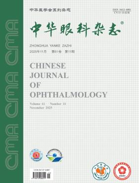
Chinese Journal of Ophthalmology
Volume 61 · Issue 11 · 2025
Chin J Ophthalmol
This issue focuses on the research progress of diagnosis and treatment of orbital diseases. The responsible editorial committee responsible for the planning, composition and review of this issue is Professor Qian Jiang.
The clinical activity score (CAS) for the thyroid-associated ophthalmopathy (TAO) evaluation has advantages such as simplicity and practicality for clinical use, but it also has limitations. Based on the formulation and development of the CAS, this article analyzes five aspects: design limitations, ethnic differences, comparison with the VISA (vision, inflammation, strabismus, and appearance/exposure) classification, controversies regarding CAS cutoff values, and the lack of objectivity in the CAS. It emphasizes that clinicians should comprehensively and dialectically evaluate the CAS by integrating the disease course, severity, and harmfulness of TAO, so as to correctly distinguish TAO stages, provide appropriate treatments, achieve favorable therapeutic effects, and ultimately improve the clinical management of TAO.
In recent years, therapeutic bandage contact lenses have been widely used in clinical practice. In 2019, the Society of Contact Lens Safety Monitoring and Visual Health of Chinese Health Association formulated expert consensus opinions on their standardized clinical application. Over the past five years, new progress has been achieved in the related research. Therefore, the society has conducted extensive discussions to attain an updated consensus on the indications, contraindications and usages of therapeutic bandage contact lenses based on the latest evidence-based medicine and clinical discoveries and experience, which may serve as a reference for clinical practitioners.
Bacterial keratitis is an acute-onset, rapidly progressive corneal infection that can lead to permanent vision loss. In recent years, shifts in the spectrum of causative pathogens and their antimicrobial resistance have created new challenges for clinical diagnosis and treatment. The Ocular Infection Group of Chinese Ophthalmologist Association has reviewed extensive domestic and international literature and combined the evidence with the expert clinical experience in China to develop a consensus statement. The document addresses the epidemiology, pathogenesis, clinical manifestations, laboratory and ancillary diagnostic methods, differential diagnosis, and therapeutic principles and options of bacterial keratitis, while underscoring the importance of individualized therapy, aiming to provide up-to-date reference and guidance for clinical practice.
To investigate the association between lipid levels and the risk of Graves′ ophthalmopathy (GO).
This study employed Mendelian randomization (MR) with a retrospective cross-sectional analysis. Summary data for lipids and GO were obtained from the genome-wide association studies (GWAS) released between 2013 and 2023, and two-sample MR was used to explore the causal relationship between lipids and GO. Inverse-variance weighting (IVW) served as the primary estimator, with MR-Egger regression and the weighted median (WM) as auxiliary methods. A series of sensitivity analyses were conducted to ensure the validity and stability of the findings. In parallel, lipid data were collected from inpatients with GO and contemporaneous healthy controls in the Department of Ophthalmology, the Third Xiangya Hospital of Central South University, from April 2022 to November 2024, and differences in lipid levels were analyzed using independent-samples t tests and non-parametric tests.
In the forward MR analysis for HDL-C, two GWAS datasets were used. Both the ieu-b-109 data (OR=0.644,95%CI=0.388-1.070,P=0.089) and the ieu-b-4844 data (OR=1.349,95%CI=0.848-2.146,P=0.206) indicated that HDL-C had no significant causal association with the risk of GO. the low-density lipoprotein cholesterol (LDL-C) was evaluated using four GWAS data sources. The ieu-a-300 data (OR=0.686,95%CI=0.352-1.335,P=0.267), the ieu-a-781 data (OR=0.467,95%CI=0.116-1.886,P=0.285), the ieu-b-110 data (OR=0.734,95%CI=0.380-1.419,P=0.358), and the ieu-b-4846 data (OR=1.311,95%CI=0.634-2.714,P=0.465) were not statistically significant, and the effect directions were inconsistent, suggesting that LDL-C had no clear causal effect on the risk of GO. Triglycerides were evaluated using five data sources. The ieu-a-783 data (OR=1.101,95%CI=0.461-2.626,P=0.829), the ieu-b-4849 data (OR=0.657,95%CI=0.203-2.128,P=0.483), the ieu-b-4850 data (OR=1.144,95%CI=0.568-2.305,P=0.707), the met-d-Total_TG data (OR=1.630,95%CI=0.309-8.600,P=0.565), and the ukb-bub35-TRIG data (OR=0.906,95%CI=0.572-1.436,P=0.674) showed no causal association between triglyceride levels and the risk of GO. the total cholesterol was evaluated using the ieu-a-782 dataset (OR=0.672,95%CI=0.103-4.399,P=0.678), which suggested no significant causal effect on the risk of GO. The effect directions of all these analyses were consistent across IVW, MR-Egger, and WM methods, suggesting that the results were relatively robust. No clear causal relationship between lipids and GO was found. In the reverse MR analysis, no causal relationship was found between GO and HDL-C, triglycerides, or total cholesterol (P>0.05). Although the ieu-b-4846 data suggested a slight inverse association between Graves′ ophthalmopathy and LDL-C (OR=0.983,95%CI=0.968-0.998,P=0.027), it was not significant after FDR correction (FDR=0.081). An observational study included 102 patients with GO, including 45 males (44.1%) and 57 females (55.9%), with an average age of 43.50 (33.00, 53.00) and a BMI of 23.55±3.50. There were 102 healthy controls, including 46 males (45.1%) and 56 females (54.9%), with an average age of 42.50 (34.00, 56.00) and a BMI of 23.20±3.67. The results showed no statistically significant differences in HDL-C (P=0.487), LDL-C (P=0.186), triglycerides (P=0.391), or total cholesterol (P=0.168) between the two groups.
Both MR and observational studies indicate that lipid levels may not be significantly associated with the risk of GO. There is a need for large sample and in-depth clinical studies in China.
To comparatively evaluate the efficacy and safety of intravitreal topotecan (IVT) versus intravitreal melphalan (IVM) combined with IVT in the treatment of vitreous seeds in retinoblastoma (RB).
Retrospective cohort study. This study collected clinical data from 34 patients with unilateral vitreous seeds in RB who were diagnosed and treated at the Department of Ophthalmology of Eye & ENT Hospital of Fudan University from December 2015 to May 2025. All patients developed refractory or recurrent vitreous seeds after receiving intravenous chemotherapy or intra-arterial chemotherapy. Baseline data, ocular characteristics and detailed treatment histories were analyzed. Patients were treated with IVT (single-drug group) or IVT combined with IVM (double-drug group), which was administered at 4-week intervals until the vitreous seeds disappeared or became stably calcified. The primary outcome measures were the control of vitreous seeds and the globe-salvage rate. The ocular and systemic adverse effects following intravitreal chemotherapy were also recorded. Statistical analyses were performed using the t test, the Mann-Whitney U test and the χ² test. The Kaplan-Meier method was used to plot globe-salvage curves and differences were compared using the log-rank test.
There were 21 (61.8%) males and 13 (38.2%) females. The median age at initial diagnosis was 29.5 (19.5, 38.0) months. Among them, 8 (23.5%) had bilateral RB, all with unilateral vitreous seeds and the affected eye received intravitreal chemotherapy and 26 (76.5%) had unilateral RB. Of the affected eyes, 15 (44.1%) were left eyes and 19 (55.9%) were right eyes. According to the International Intraocular Classification of Retinoblastoma, 13 (38.2%) eyes were classified as group E, 20 (58.8%) as group D, and 1 (3.0%) as group C. The single-drug group included 15 eyes (44.1%), with a median age at initial diagnosis of 21.0 (11.0, 30.0) months and a follow-up duration of 19.0±10.0 months. Among them, 1 eye was classified as group C, 7 eyes as group D, and 7 eyes as group E. Each affected eye received a mean of 3.1±1.1 intravitreal injections, with a total topotecan dose of 89.3±34.3 μg per eye. The double-drug group included 19 eyes (55.9%), with a median age at initial diagnosis of 33.0 (22.0, 42.0) months and a follow-up duration of 67.9±30.4 months. Among them, 13 eyes were classified as group D and 6 eyes as group E. Each affected eye received a mean of 5.0±1.7 intravitreal injections, with a total melphalan dose of 111.1±44.5 μg and a total topotecan dose of 74.7±44.0 μg per eye. During the follow-up period, vitreous seeds in all affected eyes in both groups were controlled (100%), and no orbital or systemic metastases were observed. Compared with the double-drug group, the single-drug group exhibited fewer local adverse effects. The incidence of cataract was 0/15 and 9/19, while the incidence of phthisis bulbi was 0/15 and 6/19, respectively (both P<0.05). Some patients in both groups developed retinal pigment epithelial and choroidal atrophy, vascular occlusion or optic nerve atrophy, but the differences were not statistically significant (both P>0.05). The final globe-salvage rates were 14/15 in the single-drug group and 14/19 in the double-drug group.
IVT can effectively treat vitreous seeds in RB. Compared with the regimen combining melphalan, the IVT monotherapy is associated with fewer intraocular adverse effects.
To investigate the osteogenic activity of polyetheretherketone (PEEK) modified with bone morphogenetic protein 2 (BMP2) composite hyaluronic acid hydrogel (HAH) and its feasibility for orbital fracture repair.
An experimental study was conducted from July 2022 to July 2023. PEEK scaffolds were modified with BMP2-loaded HAH to form PEEK-BMP2-HAH specimens. Rat bone marrow mesenchymal cells were transplanted into PEEK or PEEK-BMP2-HAH specimens and cultured for osteogenesis induction. Cell adhesion was observed using scanning electron microscopy (SEM). Cell proliferation was assessed using the Cell Counting Kit-8 (CCK-8). Osteogenic activity was evaluated by alkaline phosphatase (ALP) staining. Mineralized nodule formation was detected by Alizarin red staining. The relative messenger ribonucleic acid (mRNA) expression levels of Runt-related transcription factor 2 (Runx 2), osteoblast-specific transcription factor (Osterix), and osteopontin (OPN) were measured by real-time quantitative polymerase chain reaction (qPCR). A rat orbital defect model was established, and PEEK specimens (PEEK implantation group) and PEEK-BMP2-HAH specimens (PEEK-BMP2-HAH implantation group) were implanted. Bone formation was observed by hematoxylin-eosin (HE) staining, and osteocalcin (OCN) expression was detected by immunohistochemical staining.
Bone marrow mesenchymal cells adhered to and grew on the surface of both PEEK and PEEK-BMP2-HAH specimens. After 7 days of osteogenic induction, ALP staining revealed brown-black aggregated areas on PEEK discs, while PEEK-BMP2-HAH discs exhibited distinct black particles. The ALP activity in the PEEK-BMP2-HAH group [(2.36±0.18) μmol·min⁻¹·ng⁻¹ protein] was higher than that in the PEEK group [(1.00±0.18) μmol·min⁻¹·ng⁻¹ protein], with a statistically significant difference (t=12.15, P<0.001). After 14 days of osteogenic induction, Alizarin red staining demonstrated red aggregated areas on PEEK discs, whereas the PEEK-BMP2-HAH group displayed prominent reddish-brown mineralized nodules. The semi-quantitative absorbance value of Alizarin red staining in the PEEK-BMP2-HAH group (0.55±0.05) was more pronounced than that in the PEEK group (0.22±0.03), with a statistically significant difference (t=13.33, P<0.001). After 7 and 14 days of osteogenic induction, the relative mRNA expression of Runx 2 in the PEEK-BMP2-HAH group (3.25±0.22, 2.36±0.14) was significantly higher than that in the PEEK group (1.01±0.05, 1.03±0.07), with statistically significant differences (t=22.33, P<0.001; t=17.50, P<0.001). The relative mRNA expression of OPN in the PEEK-BMP2-HAH group (1.71±0.11, 2.11±0.15) was also significantly higher than that in the PEEK group (1.03±0.08, 1.04±0.10), with statistically significant differences (t=10.47, P<0.001; t=11.10, P<0.001). After 7 days of osteogenic induction, there was no significant difference in the mRNA expression of Osterix between the PEEK group (1.01±0.08) and the PEEK-BMP2-HAH group (1.12±0.09) (t=1.87, P=0.099). However, after 14 days of osteogenic induction, the relative mRNA expression of Osterix in the PEEK-BMP2-HAH group (1.77±0.17) was significantly higher than that in the PEEK group (1.02±0.06), with a statistically significant difference (t=8.95, P<0.001). HE staining of the bone defect model showed that the formation of new bone tissue was increased in the PEEK-BMP2-HAH group compared to the PEEK group. Immunohistochemical staining revealed that 12 weeks after implantation, the expression of OCN in the PEEK-BMP2-HAH group (1.98±0.25) was significantly higher than that in the PEEK group (0.98±0.11), with a statistically significant difference (t=8.07, P<0.001).
The BMP2-HAH-modified PEEK repair material exhibits stronger cell adhesion ability, proliferation activity, and osteogenic activity compared with unmodified PEEK repair material, and can promote bone tissue regeneration in a rat orbital defect model.
To observe the formation time, location, and influencing factors (including treatment regimens and systemic factors) of collateral vessels in retinal vein occlusion (RVO) patients following anti-VEGF therapy.
A retrospective cohort study was conducted among patients diagnosed with RVO at the Ophthalmology Departments of Beijing Tsinghua Changgung Hospital and Zhangjiakou Fourth Hospital between December 2017 and December 2019. Patients with macular edema were treated with anti-VEGF ranibizumab (Lucentis). Based on treatment preferences, patients were divided into three groups: no-injection group, 1+PRN group (one initial injection followed by pro re nata treatment), and 3+PRN group (three monthly injections followed by pro re nata treatment). All patients were followed up for two years, with regular collections of fundus imaging data. The primary outcome measure was the formation of retinal collateral circulation, which was assessed jointly by two senior ophthalmologists using color fundus photography and fluorescein angiography. The presence, timing, and characteristics of collateral circulation were recorded, and the effects of different treatment regimens and systemic factors were analyzed. Statistical analysis was performed using SPSS 27.0, with between-group comparisons conducted using the Chi-square tests or Fisher′s exact test. A p-value<0.05 was considered statistically significant.
A total of 157 patients (157 eyes) with RVO were included, comprising 87 cases of branch retinal vein occlusion (BRVO) and 70 cases of central retinal vein occlusion (CRVO). Among BRVO patients, 62.1% (54/87) developed collateral circulation, with a median formation time of 172.0 (253.0, 124.3) days. Among CRVO patients, 18.6% (13/70) developed collateral circulation, with a median formation time of 333.5 (369.0, 256.3) days. In BRVO patients, the rates of collateral circulation formation in the non-injection, 1+PRN, and 3+PRN groups were 9/14, 62.8% (27/43), and 90.0% (18/20), respectively. The Chi-square test indicated no statistically significant difference among the groups (χ²=5.102, P=0.078). In CRVO patients, the rates were 3/5, 19.4% (6/31), and 11.8% (4/34) in the three groups, respectively. The Fisher′s exact test revealed a statistically significant difference in collateral circulation formation rates among the treatment groups (P=0.022). The Chi-square test results showed that in BRVO patients, the presence of diabetes (χ²=2.67, P=0.10) and hypertension (χ²=0.79, P=0.37) had no statistically significant impact on the establishment of retinal collateral circulation. Similarly, in CRVO patients, the presence of diabetes (χ²=1.33, P=0.249) and hypertension (χ²=0.13, P=0.720) showed no statistically significant influence on collateral circulation formation.
Anti-VEGF therapy significantly suppressed the collateral vessel formation in patients with CRVO but had no significant effect on patients with BRVO. Systemic factors had no significant effect on the collateral formation in either group.
To achieve automated lesion detection in optical coherence tomography (OCT) images based on a lightweight convolutional neural network architecture.
This retrospective study employed deep learning to construct a lightweight lesion identification model using OCT images from Sichuan Provincial People′s Hospital alongside datasets including OCT-C8, OCT-2017, and HD-OCT of MH. Model performance evaluation comprised two stages: firstly, testing on four independent external validation sets to assess the model′s generalisability and accuracy; secondly, employing a confidence interval overlap comparison method to evaluate the classification performance of ophthalmologists at different levels of clinical experience (two per level) against the model, thereby determining the clinical experience level corresponding to the model′s medical proficiency.
The model achieved an accuracy of 93.41% (47 054/50 374) on the validation set, with an F1 score of 88.18% (44 419/50 374) and a recall rate of 86.42% (43 533/50 374). The area under the receiver operating characteristic curve was 99.31% (50 026/50 374), with precision at 91.48% (46 082/50 374) and specificity at 99.1% (49 921/50 374). For nine categories of OCT images, namely neovascularisation, posterior vitreous detachment, epiretinal membrane, macular hole, macular schisis, subretinal fluid, normal image, vitreomacular traction, and obscured image, the precision rates were 97.48% (16 873/17 310), 90.99% (3 039/3 340), 97.62% (1 325/1 358), 97.91% (2 185/2 232), 79.23% (4 961/6 262), 81.47% (1 594/1 957), 90.55% (17 744/19 596), 88.08% (237/269), and 99.95% (19 520/19 530), respectively. The model achieved an average accuracy of 89.9% on the external validation set, whereas junior physicians demonstrated an accuracy of 74% (95%CI: 61.84%~86.16%) in interpreting OCT images, and that of mid-level physicians was 88% (95%CI: 78.99%~97.01%), indicating the model′s performance approached that of mid-level physicians.
This study marks the first successful implementation of automated recognition across nine categories of OCT images. Its clinical performance has preliminarily attained the level of a mid-career physician, and it can be deployed locally within healthcare settings to enhance diagnostic efficiency and accuracy.
Four children were treated at the Ophthalmology Department of Beijing Children′s Hospital affiliated with Capital Medical University, including a female patient aged 9 years and 11 months with redness and swelling of the forehead accompanied by bilateral eyelid swelling for 3 days, a male 14-day-old infant with progressive worsening of the right eye redness and swelling accompanied by high fever, nasal congestion and a runny nose for 7 days, a boy aged 5 years and 9 months with the left eye and facial redness and swelling, black skin deposition, rupture, pus discharge and high fever for 2 days, and a 7-year-old male with the right eyelid redness and swelling accompanied by fever and acute lymphoblastic leukemia for 2 hours. The comprehensive blood routine examination showed C-reactive protein levels exceeded 140 mg/L and white blood cell counts exceeded 20×109/L in all patients. Bacterial cultures of ocular secretions or pus were mainly positive for Streptococcus pyogenes, Acinetobacter baumannii, methicillin-resistant Staphylococcus aureus, and Pseudomonas aeruginosa. Orbital imaging revealed orbital abscess formation (Chandler stage 4) in 2 patients and subperiosteal abscess and necrotizing fasciitis (Chandler stage 3) in 2 patients. According to the scoring criteria for the systemic inflammatory response syndrome, all four cases were diagnosed with severe orbital cellulitis. Systemic and ocular antibiotics were administered, along with local debridement and abscess incision and drainage. Four children recovered without any recurrence of infection. Two of them developed late-onset inner canthal deformity and eyelid abnormality, which were resolved by corrective surgery.
A 38-year-old female patient was followed up intermittently for nearly 8 years due to rhegmatogenous retinal detachment (RRD) and severe proliferative vitreoretinopathy (PVR) in the right eye. Seven years prior, the patient was diagnosed with silicone oil-dependent eye after vitrectomy and secondary cataract in the right eye; however, she declined further medical management at that time. At the latest re-evaluation (seven years after the aforementioned diagnosis), the lens that was previously affected by cataract was found to have disappeared spontaneously. The patient confirmed no surgical intervention had been received during this interval, and further examinations verified that the lens had undergone spontaneous absorption.
The patient was a 54-year-old male. Because of the gradual loss of painless vision in both eyes for 1 week, he went to the Department of Ophthalmology of Sanming No.1 Hospital. In the past, he was in good health, and he could drive normally at night 1 week ago. Within 1 week, his binocular visual function declined to the point where he could not take care of himself. General physical examination: Chest CT scan combined with enhanced examination showed pulmonary malignant tumor with hilar lymph node metastasis. Eye examination: visual acuity was 4.7 in the right eye and 4.6 in the left eye; No obvious abnormalities were observed in intraocular pressure and anterior segment; Binocular tubular field; Retinal pigment changes in both eyes, and annular pigment changes centered on the optic disc can be seen in the left eye (Quintessential Picture 1). Coherence light tomography examination: except for the very small area in the center of the macula, the rest of the outer retinal structure disappeared (Quintessential Picture 2). Fundus autofluorescence imaging examination: extensive diffuse strong fluorescence in the right eye, strong fluorescence around the optic disc and posterior pole in the left eye, and patchy weak fluorescence around the optic disc and macular area in both eyes (Essence Picture 3). Serum retinal antibody test: anti-recoverin antibody IgG positive. Clinical diagnosis: paraneoplastic autoimmune retinopathy. He was given oral glucocorticoid therapy and transferred to the respiratory department for diagnosis and treatment. After 2 weeks of treatment, ophthalmic review showed that the visual acuity of the right eye was light sensing, but the left eye had no light sensing.
Polypoidal choroidal vasculopathy (PCV) is a common subtype of neovascular age-related macular degeneration (nAMD) in Asian populations. Clinical follow-up has revealed varying degrees of activity in PCV lesions. Studies have shown that multimodal retinal imaging techniques can be used to assess the activity of PCV lesions. Abnormal branching vascular networks, polypoidal lesions, and choroidal-related features are important biological markers for predicting lesion activity. Additionally, demographic characteristics and genetic traits of patients may also serve as relevant factors for assessing the activity of PCV lesions. This review summarizes recent updates in the understanding of PCV lesion activity based on multimodal retinal imaging studies and their impact on clinical ophthalmic practice. It also explores the potential applications and prospects of lesion activity characteristics in the treatment and follow-up of PCV, aiming to provide reference for clinicians in assessing and determining the activity of PCV lesions and developing personalized treatment and follow-up plans.
Functional visual disorder (FVD) belongs to functional neurological disorder (FND), and clinical examination and testing are not consistent with clinical ophthalmic or neurological disease.The major clinical manifestations are visual acuity loss and visual field loss.FVD has a serious impact on patients′ daily life, and it is difficult to distinguish from other visual disorders by its clinical manifestations. Some patients receive unnecessary treatments, so early diagnosis and accurate treatment are particularly important. This article reviews the clinical characteristics, pathological mechanism, diagnosis and differential diagnosis, auxiliary examination, management and prognosis of FVD in order to improve clinical diagnostic and therapeutic rates and patient prognosis.
Nanophthalmos is a rare congenital ocular disorder characterized by the reduction of both the anterior and posterior segments of the eye, occurring without associated ocular malformations. This condition predisposes individuals to secondary angle-closure glaucoma, a leading cause of vision loss. Due to the rarity of nanophthalmos, current research is limited, primarily focusing on its clinical classification, genetic underpinnings, and complications. This review synthesizes existing literature on the pathogenesis, epidemiology, ocular parameter changes related to glaucoma, and surgical interventions for secondary glaucoma in nanophthalmic patients, emphasizing studies from the past five years to enhance understanding in clinical research and management.
Orbital surgery needs to complete delicate operations in a narrow space and limited visual field on the premise of avoiding damage to important tissues in the orbit. The surgery is characterized by high difficulty, high risk and many complications. The application of digital medical technologies represented by three-dimensional (3D) modeling, additive manufacturing (also known as 3D printing), surgical navigation, extended reality, artificial intelligence, and surgical robots in orbital surgery has improved the safety and accuracy of surgery, and pushed orbital surgery to the road of individualization, precision and intelligence. This article will card and discuss this, in order to provide a brick for the long-term development of orbital surgery.
The Cataract and Refractive Surgery Group of Ophthalmology Branch of Chinese Medical Association and the Cataract Group of Ophthalmologists Branch of Chinese Medical Doctors Association jointly updated and formulated the Expert Consensus on Clinical Application of Presbyopia Correction Intraocular Lens in China (2025) based on the latest evidence-based medical evidence. In order to help ophthalmologists deeply understand the content of the consensus, the indications, contraindications, preoperative evaluation and decision-making of presbyopia corrective intraocular lens are now interpreted and explained.
Recently, the Cataract and Refractive Surgery Group of Ophthalmology Branch of Chinese Medical Association and the Cataract Group of Ophthalmologists Branch of Chinese Medical Doctors Association jointly formulated the Expert Consensus on Clinical Application of Presbyopia Correction Intraocular Lens in China (2025). In order to help the majority of ophthalmologists to further understand the formation basis of consensus opinions and clinical practice schemes, this article interpreted the key technologies, adverse reactions and management, doctor-patient communication related to the operation in presbyopia corrective intraocular lens implantation surgery, and summarized the latest progress of presbyopia corrective surgery.
CURRENT ISSUE