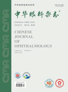
Chinese Journal of Ophthalmology
Volume 61 · Issue 04 · 2025
Chin J Ophthalmol
This issue focuses on the research progress of ocular biomechanics. Ocular biomechanics plays an important role in the pathogenesis, diagnosis and treatment of many ophthalmic diseases. It not only involves the mechanical interaction between intraocular pressure and eyeball wall, but also deeply affects the morphology and function of ocular structures such as cornea and lens. The occurrence and development of many eye diseases, such as corneal dilatation in keratoconus, intraocular pressure regulation in glaucoma and lens deformation in myopia, are closely related to the changes of ocular biomechanics. In-depth research on ocular biomechanics will help to reveal the essence of disease and provide new theoretical basis and technical means for clinical diagnosis and treatment. The responsible editorial committee responsible for the planning, composition and review of this issue is Professor Wang Yan. In this issue, she published an expert review article "Emphasizing the Role of Biomechanics in the Pathogenesis and Diagnosis and Treatment of Eye Diseases", which deeply discussed how biomechanical factors participate in the occurrence and development of eye diseases, and analyzed the current application status and challenges of biomechanics in clinical diagnosis and treatment of ophthalmology. It is pointed out that many eye diseases are closely related to biomechanics, such as force imbalance of keratoconus, changes of intraocular pressure in primary glaucoma and corneal dilation after refractive surgery. By combining biomechanics with other disciplinary knowledge, it is expected to provide new ideas and methods for the diagnosis and treatment of difficult eye diseases, and promote the development of eye health.
Biomechanics is an interdisciplinary field that studies the intrinsic relationship between biology and mechanics. It plays a crucial role in the occurrence, development, diagnosis, and treatment of diseases. In ophthalmology, biomechanics is particularly relevant to the mechanisms of ocular growth, development, pathological changes, injury repair, disease progression, and the diagnostic and therapeutic approaches associated with ocular diseases. This article addresses three key aspects: the insufficient attention given to biomechanics in ophthalmology, the potential of biomechanics as a critical breakthrough in overcoming clinical challenges in ophthalmic diagnosis and treatment, and the need to gradually establish a new diagnostic and therapeutic paradigm that incorporates biomechanics. We emphasize the importance of biomechanics in the pathogenesis and diagnosis of ocular diseases, highlight the urgent issues, and advocate for greater focus on the role of biomechanics to guide clinical practice.
Glaucoma stands as the leading cause of irreversible blindness globally, yet its pathogenesis remains incompletely understood. With the advancements in biomechanical detection technologies, in-vivo biomechanical research on the eyeball has become feasible. An increasing number of researchers have turned their attention to the intricate relationship between the biomechanical properties of the cornea and the onset and progression of glaucoma. This article delves into four crucial aspects: grasping the concepts and detection techniques of corneal biomechanics, exploring the correlation between corneal biomechanics and glaucoma, investigating the influencing factors of corneal biomechanics in glaucoma cases, and understanding the application of corneal biomechanics-corrected intraocular pressure in glaucoma diagnosis and treatment. It emphasizes the need to comprehensively recognize the reference value of corneal biomechanical parameters in glaucoma diagnosis and treatment. The authors aim to collaborate with fellow ophthalmologists in related research, with the ultimate goal of enhancing the diagnostic and therapeutic standards for glaucoma.
To investigate the biomechanical properties of the lens and the changes before and after cycloplegia in the myopic population in China, and to analyze the possible influencing factors.
This is a cross-sectional study. Consecutively, 159 myopic patients (159 right eyes) who underwent the Brillouin Optical Scanning System examination from November 2023 to May 2024 at Tianjin Eye Hospital were recruited. Among them, 82 were male and 77 were female, with an age of 21 (18, 24) years. The patients were classified into low, intermediate, and high myopia groups according to the equivalent spherical lens power. The lens biomechanical parameters, as well as ocular refractive and morphological parameters, were measured, including the width of top plateau, the width of bottom plateau, the longitudinal modulus of nucleus (LMN), the longitudinal modulus of cortex (LMC), the axial length of the eye, lens thickness, anterior radius of curvature, posterior radius of curvature, and lens density. The differences in lens parameters before and after cycloplegia were compared. Univariate and multivariate linear regression analyses were utilized to assess the factors associated with the LMN before and after cycloplegia.
There were 46, 59, and 54 patients in the low, intermediate, and high myopia groups respectively. There were no statistically significant differences in age or gender distribution among the three groups (all P>0.05). The LMN of the three groups of low, intermediate, and high myopia were 3.383 (3.361, 3.411), 3.401 (3.378, 3.416), and 3.415 (3.395, 3.422) GPa, respectively, and the LMN was higher in the high myopia group; the difference in the LMN among the three groups was statistically significant (P<0.001). The difference in the LMC of the lens cortex was not statistically significant among the 3 groups of low, intermediate, and high myopia (P>0.05). After ciliary muscle paralysis, the LMN decreased slightly compared with that before paralysis [3.395 (3.371, 3.414) and 3.405 (3.379, 3.417) GPa, P<0.05]. The results of multifactorial linear regression analysis showed that LMN was proportional to refractive error both before and after ciliary muscle paralysis, and the higher the myopia, the greater the LMN (all P<0.05). The greater the lens optical density, the smaller the LMN (all P<0.05). Gender was also associated with LMN (all P<0.05).
In young myopic patients, LMN increased with the increase of myopia degree and was higher in highly myopic patients, suggesting that the biomechanical properties of the myopic lens may be altered. Moreover, there were differences in LMN before and after ciliary muscle paralysis, suggesting that accommodation may affect the mechanical properties of the lens. Gender and lens optical density are also relevant factors affecting LMN.
To compare the 1-year clinical efficacy differences of high-ultraviolet energy epithelium-removing accelerated corneal collagen cross-linking (A-CXL) in the treatment of primary keratoconus in juveniles and adults.
This is a retrospective cohort study. The data of 72 patients (110 eyes) with keratoconus who received high-ultraviolet energy epithelium-removing A-CXL in the Ophthalmology Center of Xiangya Hospital, Central South University from August 2022 to December 2023 were collected. There were 52 male patients and 20 female patients, with aged (20.3±5.5) years. According to the age, they were divided into a juvenile group (≤18 years old, 35 cases, 56 eyes) and an adult group (>18 years old, 37 cases, 54 eyes). The visual acuity (uncorrected distant visual acuity, best corrected visual acuity), corneal endothelial parameters [corneal endothelial cell density, coefficient of variation of corneal endothelial cell area (CV), hexagonal cell ratio], morphological parameters [flat corneal curvature, steep corneal curvature, maximum corneal curvature (Kmax), central corneal thickness, thinnest point corneal thickness, etc.] and biomechanical parameters [stress-strain index (SSI), horizontal Ambrósio-related thickness (ARTh), etc.] before surgery and at 1, 3, and 12 months after surgery were compared between the two groups.
The best corrected visual acuity of the juvenile group at 1, 3, and 12 months after surgery was changed from 0.09±0.21 before surgery to 0.20±0.23, 0.13±0.22, and 0.14±0.22 respectively (all P<0.05). The best corrected visual acuity of the adult group was only changed from 0.16±0.29 to 0.24±0.30 at 1 month after surgery (P<0.05). The CV of the juvenile group at 1 month after surgery was 29.3%±2.1%, which was significantly higher than 28.4%±2.3% before surgery (P<0.05), while the CV of the adult group remained stable (P>0.05). At 1 month after surgery, the Kmax of the juvenile group increased from (53.85±6.56) D before surgery to (55.00±6.93) D, and that of the adult group increased from (54.13±10.02) D to (56.41±8.55) D (both P<0.05), and both returned to the baseline level at 3 months after surgery. The absolute values of the changes in Kmax of the adult group at 3 months and 12 months after surgery compared with the preoperative baseline values were (1.06±2.49) D and (1.52±2.62) D respectively, which were significantly higher than (0.01±2.37) D and (0.43±2.57) D of the juvenile group (both P<0.05). Biomechanical analysis showed that the SSI of the adult group at 3 months after surgery was 0.79±0.16, which was significantly higher than 0.72±0.13 of the juvenile group (P<0.05), while the ARTh was (187.3±81.8) μm, which was lower than (235.0±117.0) μm of the juvenile group (P<0.05). By 12 months after surgery, the SSI of the adult group was still higher than that of the juvenile group (P<0.05).
Within 1 year after high-energy A-CXL, the improvement of corneal stiffness is more significant and the changes in corneal morphology are more obvious in adult patients, suggesting that the short-term efficacy of this surgical procedure for adult keratoconus is better than that for juvenile patients.
To investigate age-related dynamics of refractive compensation for axial elongation in children aged 6-9 years, and examine the modulating effects of follow-up duration, baseline age, and refractive status.
This prospective cohort study analyzed data from the Shanghai Time Outside to Reduce Myopia Trial (2016—2020), including 1 805 non-myopic or low-myopic children [mean age (6.77±0.35) years; 52.0% male]. Quantile regression analysis characterized spherical equivalent changes (ΔSE) per millimeter of axial elongation (ΔAL) across different observation periods (1-4 years). Mixed-effects models evaluated moderating factors including age and baseline refractive status.
Axial elongation demonstrated strong positive correlations with refractive changes across all follow-up durations (r:0.79-0.91, P<0.001). The median refractive compensation per millimeter axial elongation exhibited significant temporal attenuation: 2.06 D (1.84-2.31 D) at 1-year follow-up decreasing to 1.50 D (1.25-1.75 D) over 3-4 years (P-trend<0.001). Each additional baseline year reduced compensation magnitude by 0.19-0.31 D (P<0.05), with 6-year-olds showing 30.1% reduction in compensation efficiency (2.06 -1.44 D) over 4 years. Non-myopic eyes demonstrated 28.0% lower baseline compensation (1.44 D vs. 2.00 D, P<0.001) with slower annual attenuation (0.03 D vs. 0.19 D) compared to myopic eyes.
Pediatric refractive compensation follows a dual attenuation pattern modulated by both age and time, with more pronounced compensation loss in myopic eyes. These findings emphasize the necessity for dynamic adjustment of axial length-to-refraction conversion formulae incorporating follow-up duration, baseline age, and refractive status to enhance myopia progression risk assessment.
To investigate the developmental characteristics of visual acuity in C57BL/6J mice and to evaluate the consistency and reliability of different measurement methods.
This was an experimental study. One hundred and seventy-two C57BL/6J mice were divided into eight groups based on postnatal days (P): P13/14 (first day of eye opening,18 mice), P15 (11 mice), P16 (10 mice), P18 (21 mice), P21 (onset of the critical period for visual development, 29 mice), P28 (peak of the critical period, 28 mice), P35 (end of the critical period, 25 mice), and P60 (adulthood, 30 mice). Visual acuity was assessed using three methods: visual evoked potentials (VEP), the optomotor reflex (OMR) task, and the visual water task (VWT). Statistical analyses were conducted using the least significant difference t-test, one-way ANOVA, and independent sample t-tests.
VEP measurements of monocular visual acuity showed that the visual acuity in the P13/14 group was (0.20±0.06) cycles per degree (cpd), and increased to (0.44±0.08) cpd in the P21 group and (0.52±0.06) cpd in the P35 group, with significant differences between groups (all P<0.05). The visual acuity in the P60 group was (0.56±0.05) cpd, which was not significantly different from that in the P35 group (P>0.05). For binocular visual acuity assessed by the OMR task, the P13/14 group had a visual acuity of (0.11±0.06) cpd, which increased to (0.20±0.07) cpd in the P15 group, (0.35±0.05) cpd in the P16 group, (0.40±0.05) cpd in the P18 group, and (0.47±0.05) cpd in the P21 group, with significant differences between groups (all P<0.05). The visual acuity in the P60 group was (0.50±0.05) cpd, which was not significantly different from that in the P21 group (P>0.05). The binocular visual acuity assessed by the VWT in the P60 group was (0.50±0.03) cpd. There was no significant difference in visual acuity measured using the three methods in the P60 group (P>0.05).
Visual acuity in C57BL/6J mice increased rapidly from the first day of eye opening to the onset of the critical period for visual development, followed by a slower increase until adulthood. In adult C57BL/6J mice, it ranged from 0.50 to 0.56 cpd, as measured using the VEP, OMR task, and VWT, which can yield comparable and reliable results.
To systematically evaluate the predictive accuracy of different intraocular lens (IOL) power calculation formulas in cataract patients with short axial length (≤22.5 mm).
This is a meta-analysis. Databases including PubMed, Web of Science, Cochrane Library, Embase, and CNKI (from the establishment of the database to August 2023) were searched, and case-control studies comparing≥2 IOL formulas were included. The literature involves 12 intraocular lens (IOL) power calculation formulas, including Barrett Universal Ⅱ, EVO, Haigis, Hill RBF, Hoffer Q, Holladay 1, Kane, Olsen, PEARL DGS, SRK/T, T2, and VRF. The primary outcome measures were cases with prediction errors within the ranges of±0.25 D,±0.50 D, and ±1.00 D.Two researchers independently screened the literature, extracted data, and evaluated the risk of bias (using the QUADAS-2 tool). Traditional meta-analysis (fixed/random-effect model) was performed using RevMan 5.3, and network meta-analysis was carried out using Stata 14. The proportion of predictive errors within the ranges of ±0.50 D and ±1.00 D were the main indicators. The odds ratio (OR) and its 95% confidence interval (CI) were used to compare and evaluate the predictive error situations of different formulas.
A total of 8 studies (785 eyes) were included. Traditional meta-analysis showed that within the ±1.00 D range, Hoffer Q (93.4%; 648/694) and Kane (95.7%; 426/445) were significantly better than SRK/T (OR=1.50, 95%CI: 1.01-2.25, P=0.020; OR=2.00, 1.73-3.43, P=0.007). Kane was signifficantly better than T2 (OR=2.09, 1.15-3.80, P=0.020); within the ±0.50 D range, PEARL-DGS was better than SRK/T (OR=1.34, 1.00-1.80, P=0.050); within the ±0.25 D range, EVO (46.1%; 94/204) had the highest proportion and was significantly better than Hoffer Q (OR=0.62, 0.41-0.91, P=0.020). Network meta-analysis (ranked by the area under the cumulative ranking curve) indicated that EVO (ranked first within ±0.50 D) and Kane (ranked first within ±1.00 D) had the best overall performance, and SRK/T was the worst in all ranges (P<0.05).
Among the traditional optical refraction formulas, Haigis and Hoffer Q have good accuracy, and SRK/T is not recommended; the new-generation formulas (EVO, Kane) have better prediction accuracy in patients with short axial length and are recommended for clinical priority selection.
The patient was a 38-year-old female. He went to the Department of Ophthalmology of Peking Union Medical College Hospital because of painless blurred vision in both eyes for 2 weeks. The patient developed fever symptoms after intravenous drug infusion 2 weeks ago, followed by paroxysmal blurred vision in both eyes, and was given symptomatic treatment. After the fever symptoms were relieved, the attacks recurred, and the conscious visual acuity further decreased. Systemic physical examination: Candida albicans (+), blood 1,3-beta-D glucan concentration (G test) was 772.1 pg/ml in blood culture examination. Eye examination: the best corrected visual acuity was 0.25 for the right eye and 0.8 for the left eye; There were no obvious abnormalities in intraocular pressure and anterior segment in both eyes; Mild inflammatory opacity of the vitreous body in both eyes, with scattered yellowish-white lesions in the posterior pole retina (Quintessential Picture 1). Coherence optical tomography examination: massive hyperreflective signals in each layer of the retina in both eyes (Quintessential Picture 2). Clinical diagnosis: fungal chorioretinitis in both eyes; Candida bacteremia.
The occurrence and development of biological tissue diseases are closely related to their biomechanical properties. In recent years, significant achievements have been made in the research on the biomechanical properties of the cornea and sclera, and the research on the association between the biomechanics of the posterior eye and eye diseases has also been continuously advanced. This review systematically collates the research on the biomechanical characteristics of the retina, Bruch′s membrane-choroid complex (BMCC), and optic nerve head. The retina is anisotropic, with the elastic modulus increasing from the inner layer to the photosensitive layer. The internal limiting membrane has high mechanical strength and stiffness, and these properties change with age. The stiffness of the BMCC increases with strain, and its biomechanical changes are closely linked to aging and age-related diseases. The stress-strain pattern of the optic nerve head is affected by multiple factors and plays a crucial role in diseases such as glaucoma. These biomechanical properties are of great significance in the pathogenesis of diseases such as age-related macular degeneration, diabetic retinopathy, and glaucomatous optic neuropathy, and are also widely applied in the optimization of clinical surgeries, disease monitoring, and diagnosis. However, currently, the research on the biomechanics of the posterior segment of the eye is still in its infancy. The mechanical regulation mechanisms of multiple factors such as age, intraocular pressure, blood flow, and axial length have not been fully elucidated, and aspects such as matrix protein remodeling and post-translational modifications also require in-depth research.
Refractive error is a significant factor contributing to visual impairment, imposing a relatively large burden on the social economy. Although refractive surgery is an important corrective method, it faces challenges in clinical practice, such as precise preoperative screening, personalized surgical plan design, and prevention of postoperative complications. This study focuses on the application of machine learning in the field of refractive surgery. Through a comprehensive analysis of relevant literature, it is found that machine learning plays a positive role in multiple key aspects. In preoperative screening, it can effectively improve the accuracy of keratoconus screening and assist in precisely selecting surgical candidates and determining surgical methods. During surgical design, it can optimize the plans for corneal refractive surgery and implantable Collamer lens implantation, enhancing the predictability of surgeries. In postoperative evaluation and prediction, it helps to assess surgical outcomes, identify high-risk patients for refractive regression, and assist in calculating the power of intraocular lenses. However, machine learning has limitations in practical applications, such as the "black box" nature of algorithms, uneven data quality, and lack of multimodal data integration. By systematically reviewing its application status and limitations, this review hopes to provide references for subsequent research, help overcome difficulties, and promote the more in-depth and rational application of machine learning in the field of refractive surgery, thereby improving the overall level of refractive surgery.
Corneal neovascularization, a severe pathologic change of the ocular surface, is usually caused by complex pathogeneses, and leads to severe vision damage and eventually corneal blindness. The applications of anti-inflammatory and anti-angiogenic drugs as well as keratoplasty have proved to have certain therapeutic effects. However, there are still numerous barriers that have not yet been fully overcome, prompting a trend towards treatment with multidisciplinary techniques. In this review, we summarize the latest research progress and give an overview of the techniques and strategies for treatment of corneal neovascularization in recent years based on the new mechanistic findings, aiming to improve the therapeutic efficiency and precision.
As the global prevalence of myopia continues to rise, effective myopia prevention and control among children and adolescents is particularly important. Adjustment and binocular vision function are closely related to the progression and prevention and control of myopia, and are also important indicators for comprehensively evaluating the effectiveness of myopia prevention and control. This paper systematically discusses the adjustment and binocular vision function in myopia prevention and control from four aspects: adjustment and binocular vision function evaluation, adjustment and binocular vision function in the occurrence and progression of myopia, adjustment and binocular vision function in myopia prevention and control and its influence on myopia prevention and control effect, adjustment and binocular vision function training of myopic children, aiming at improving the effectiveness of myopia prevention and control and further promoting the development of personalized precision myopia prevention and control.
CURRENT ISSUE