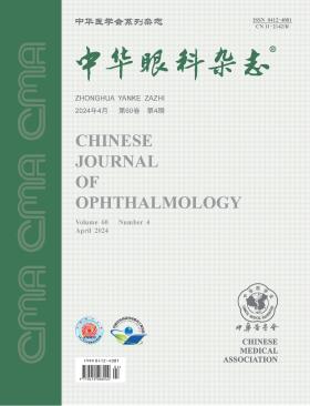
Chinese Journal of Ophthalmology
Volume 60 · Issue 04 · 2024
Chin J Ophthalmol
This issue focuses on the research progress of children's vision problems. The responsible editorial committee responsible for the planning, composition and review of this issue is Professor Li Li.
The incidence of myopia is high in China. The proportion of high myopia is also high in the myopic population. High myopia is associated with multiple fundus changes, among which the neuropathic damage is usually ignored, and thus there has been limited clinical research on the pathogenesis, standard follow-up and effective treatment of optic neuropathy in high myopia. This article focuses on the types of high myopia-associated neuropathic changes, the quantitive imaging of neuropathic damage, and the need of relevant cohort studies and pathogenesis research, aiming to attract more attention to optic neuropathic changes in high myopia.
Infantile esotropia is a common ophthalmic disease in children. A lot of clinical and basic research evidence suggests that early surgery enhances sensory and ocular motor development. However, the proper timing of surgery has been debated for decades. In addition, there is more likely instability of deviation in the preoperative evaluation of infants, and even if the patient achieved alignment after surgery, the defects in binocular vision may accompany for a lifetime. This article analyzes the difficulties and key points of early intervention for infantile esotropia, aiming to provide scientific ideas for the early treatment of children with infantile esotropia in China.
The International Myopia Institute introduced the concept of "pre-myopia" in 2019, defining it as children with refractive error ≤+0.75 D and >-0.50 D. By considering baseline refractive error, age, and other quantifiable risk factors, there is a significant likelihood that individuals falling into this category may develop myopia in the future. Therefore, it is deemed worthwhile to implement preventative intervention measures.This article delves into the epidemiology of premyopia in children and adolescents, the criteria for determining pre-myopia, the characteristics of refractive changes, and the existing evidence gaps in pre-myopia control technology. Furthermore, it explores the role and advantages of implementing pre-myopia control to enhance efforts in preventing and managing myopia. The paper highlights the essential value and future trajectory of pre-myopia control in the comprehensive management of myopia. The ultimate goal is to foster collaboration with professionals, aiming to discuss innovative strategies for effectively preventing and managing the onset and progression of myopia.
Asthenopia is a group of eye discomfort syndromes with complex clinical manifestations, accompanied with systemic symptoms or not. It is caused by diverse influencing factors and occurs upon overuse of the eyes. Over the past decade, significant changes have appeared in the etiology, clinical manifestation and treatment of asthenopia with great changes in the society and lifestyles. The Chinese Optometric Association of Chinese Ophthalmological Society and the Optometry Group of Chinese Ophthalmologist Association have organized nationwide experts in the field to thoroughly discuss the latest researches on asthenopia. Consensus opinions have been formed on the causes, influencing factors, clinical manifestations, diagnosis and treatment of asthenopia, following the definition of asthenopia in the International Classification of Diseases, so as to provide guidance for the clinical diagnosis and treatment of asthenopia.
To investigate the influence of corneal e-value on the effectiveness of orthokeratology in controlling myopia in children and adolescents.
A retrospective cohort study was conducted, involving the data from 1 563 myopic patients (1 563 eyes) who underwent orthokeratology at the Affiliated Eye Hospital of Shandong University of Traditional Chinese Medicine from June 2015 to August 2021 and adhered to lens wear for at least 2 years. The cohort consisted of 737 males and 826 females with an average age of (10.84±2.13) years. Based on corneal e-value parameters obtained from corneal topography, patients were categorized into a low e-value group (n=425) and a high e-value group (n=1 138). Data on gender, age, parental myopia history, and baseline measures such as spherical equivalent (SE), axial length, and corneal e-value were collected. Differences in axial length change and corneal fluorescein staining rates were compared between the two groups at 1 and 2 years after the start of lens wear. A generalized linear mixed model was established with axial length change as the dependent variable to analyze the correlation between axial length change and baseline corneal e-value.
The initial age of the 1 563 myopic patients was (10.84±2.13) years, with a baseline SE of (-3.05±1.30) D. After 1 year of lens wear, the axial length change was (0.20±0.19) mm in the low e-value group and (0.24±0.20) mm in the high e-value group. After 2 years, the changes were (0.38±0.25) mm and (0.43±0.27) mm, respectively, with statistically significant differences (all P<0.05). The incidence of corneal staining after 1 year of lens wear was 9.2% (39/425) in the low e-value group and 14.1% (160/1 138) in the high e-value group. After 2 years, the rates were 15.8% (67/425) and 21.8% (248/1 138), respectively, with statistically significant differences (all P<0.05). After adjusting for parental myopia history, age, SE, and baseline axial length, the baseline corneal e-value was positively correlated with axial length change at 1 and 2 years after lens wear (all P<0.05).
Corneal e-value is an independent factor influencing the effectiveness of orthokeratology in controlling myopia. A smaller corneal e-value is associated with slower axial length growth after orthokeratology, indicating better control of myopia in treated eyes.
To explore the distribution characteristics of peripheral retinal defocus in children and adolescents.
This cross-sectional study included 500 individuals aged 3 to 18 years, who visited the People's Hospital of Lincang, the First Affiliated Hospital of Dali University and Dali Ophthalmology Hospital between January and December 2021. Data of the right eye of each participant was analyzed. There were 226 males (45.20%) and 274 females (54.80%), with an average age of (10.79±3.79) years. All participants underwent post-cycloplegic refraction, optical biometry, and intraocular pressure measurement to obtain spherical equivalent, average corneal curvature, axial length, and intraocular pressure. Multispectral refraction topography was performed to obtain topographic maps and values at various field angles and orientations of peripheral retinal defocus. Based on multispectral refraction topography, peripheral retinal defocus values were categorized as crater type, hemilateral upturn type, saddle type, and relatively flat type. The distribution of different refractive states was analyzed.
The spherical equivalent of the 500 participants was(-1.51±2.61) D, axial length was (24.10±1.28) mm, and average corneal curvature was (43.20±1.22) D. Among the 500 eyes, 382 exhibited hyperopic peripheral retinal defocus values, with 316 eyes (82.72%) being myopic. Myopic peripheral retinal defocus values were observed in 118 eyes, with 15 eyes (12.72%) being myopic. Among different types of peripheral retinal defocus values, 112 eyes (22.4%) exhibited a crater type, 153 eyes (30.6%) exhibited a hemilateral upturn type, 107 eyes (21.4%) exhibited a saddle type, and 128 eyes (25.6%) exhibited a flat type. The proportion of myopia was 82.14% (92 eyes), 69.28% (106 eyes), 60.75% (65 eyes), and 3.90% (5 eyes), respectively. The peripheral retinal defocus values at 15°, 30°, and 45° were (0.01±0.08) D, (0.06±0.21) D, and (0.20±0.37) D, respectively. The peripheral retinal defocus values at temporal, inferior, nasal, and superior locations were (0.58±0.69) D, (0.52±0.63) D, (0.21±0.64) D, and (-0.26±0.67) D, respectively. Notably, the superior primarily manifested as myopic, while the others were predominantly hyperopic.
Approximately three-fourths of children and adolescents exhibit hyperopic peripheral retinal defocus values, with a higher prevalence of myopia in this subgroup. The hyperopia peripheral retinal defocus value increases with the distance from the retina to the macula. The peripheral retinal defocus values between superior and inferior, nasal and temporal locations are asymmetrical, with the temporal hyperopic peripheral retinal defocus value being most prominent and the superior myopic peripheral retinal defocus value being most evident.
The aim of this paper is to compare the refractive correction effects of rigid gas permeable contact lenses (RGPCL) and spectacle correction in children with aphakia after congenital cataract surgery.
This was a prospective non-randomized controlled trial. Children with aphakic eyes after congenital cataract surgery, who underwent vision correction in the Strabismus and Pediatric Ophthalmology Clinic of Beijing Tongren Hospital affiliated with Capital Medical University from April 2012 to November 2019, were continuously collected. Those who voluntarily chose to wear RGPCL for refractive correction were included in the experimental group. Patients with monocular disease were in trial group 1, and patients with binocular disease were in trial group 2. Patients who chose to wear frame glasses for refractive correction were included in the control group. Patients with monocular disease were in control group 1, and patients with binocular disease were in control group 2. Regional origin, medical history, and family information were collected at the first diagnosis. During the follow-up, adverse reactions occurring during the process of wearing glasses were recorded. The Teller acuity card was used for visual examination to obtain the best-corrected visual acuity and convert it into the logarithm of the minimum resolution angle. The degree of nystagmus was determined according to the amplitude and frequency of nystagmus. Treatment cost, treatment compliance, and the reasons for adopting or not adopting RGPCL were analyzed through a questionnaire completed by the parents of children with RGPCL.
A total of 203 children (344 eyes) who underwent congenital cataract surgery were included, including 124 males (210 eyes) and 79 females (134 eyes). The age range was 3 to 36 months. There were 28 cases in the experimental group, including 19 cases in trial group 1 and 9 cases in trial group 2. There were 175 cases in the control group, including 43 cases in control group 1 and 132 cases in control group 2. Except for 6 months of age, the visual acuity of the experimental group was better than that of the control group, and the differences were statistically significant (P<0.05). The visual acuity of children in trial group 1 was better than that of children in control group 1 at the same age. Among them, at 12 months of age [1.54 (1.27, 1.97), 1.84 (0.97, 2.12)], 18 months of age [1.27 (0.97, 1.84), 1.84 (0.97, 2.12)], 24 months of age [1.54 (1.27, 1.84), 1.84 (0.97, 2.12)], and 30 months old [0.97 (0.66, 1.27), 1.54 (0.66, 2.12)], the difference was statistically significant (P<0.001). The visual acuity of children in trial group 2 was better than that in control group 2 at the same age. Among them, at 18 months old [1.27 (0.97, 1.54), 1.27 (0.66, 2.12)], 24 months old [0.97 (0.66, 1.27), 1.27 (0.66, 2.12)], and 30 months old [1.27 (0.66, 2.12)], the difference was statistically significant (P<0.05). The remission rate of nystagmus in the experimental group was 8/9 (8 cases), the remission rate of nystagmus in the control group was 34.40% (32 cases), and the exacerbation rate was 29.03% (27 cases). The average annual cost of the experimental group was 25 125 yuan, and that of the control group was 2 511 yuan.
RGPCL is a well-tolerated, safe, and effective treatment for infants and young children. The visual acuity and degree of nystagmus were significantly improved in children who wore RGPCL for aphakia refractive correction after congenital cataract surgery compared with spectacle correction.
To investigate the differences in reading efficiency and visual fatigue between the use of augmented reality (AR) glasses and laptops.
A prospective self-controlled study was conducted. Healthy students from Capital Medical University who frequently engaged in long-term near work and used laptops and other digital display devices were recruited as subjects at Beijing Tongren Hospital, Capital Medical University between November 1 and November 15, 2023. LogMAR visual acuity, visual functions (accommodation, convergence, and fusion), and visual fatigue scores (Likert visual fatigue scale) of the participants were assessed. The order of using the laptop and AR glasses for each participant was determined by a coin toss. Reading efficiency (reading speed and error rate multiplied by the detection rate of incorrect numbers) with different devices for 10 minutes at the same time on different dates and visual fatigue scores after watching a 20-minute video were measured. Statistical analyses were performed using paired t-tests and Wilcoxon signed-rank tests.
A total of 20 eligible subjects were included, comprising 7 males and 13 females, with a mean age of (25.45±2.27) years. There was no significant change in binocular visual acuity before and after using AR glasses and laptops (both P>0.05). The reading speed and reading efficiency of using AR glasses [(34.03±9.25) and (29.19±7.62) digits/min, respectively] were significantly lower than those of using laptops [(39.43±10.36) and (35.67±9.87) digits/min, respectively] (t=4.36, P<0.001), while the difference in error detection rate was not statistically significant (t=1.29, P=0.213). There was no statistically significant difference in visual fatigue scores before watching videos with the two devices (Z=-0.71, P=0.480). However, the visual fatigue score after watching videos with AR glasses [(20.55±5.04) points] was significantly higher than that with laptops [16.50 (13.00, 19.75) points] (Z=-2.85, P=0.004). The visual fatigue scores after watching videos with both devices were significantly higher than before (P<0.05), with a more significant increase observed with AR glasses [(6.05±3.50) points] (Z=-3.41, P<0.001).
Compared with using laptops, the reading speed and efficiency were lower, and the visual fatigue was more pronounced with the use of AR glasses at the current technical level. Further optimization and improvement of AR glasses are warranted.
To analyze the current research status of uveitis in China.
It was a bibliometric analysis study. Using search formulas covering uveitis and its multiple subtypes, uveitis-related literature in English with publication dates from 2013 to 2022 was retrieved in Web of Science core databases through certain search strategies. This study used the latent Dirichlet allocation (LDA) algorithm to build topic models and analyzed the trends of research topics in recent years. Bibliometric analysis was used to analyze and visualize the bibliometric indicators (e.g., number of publications, citations, and H-index) of the included literature using tools such as VOSviewer software.
Over the past decade, China has published 1 657 papers on uveitis, ranking second globally. However, there is still room for improvement in terms of the H-index (58) and citation (12.28 per publication). Countries such as the USA (43.04%) and the United Kingdom (62.54%) were engaged in more international collaboration. We identified ten optimal LDA topics for uveitis literature in China such as immunotherapy, Behçet′s disease, and Vogt-Koyanagi-Harada syndrome. Research on uveitis in China was mostly published in Ocular Immunology and Inflammation (92).
China has made remarkable progress in uveitis research. Nonetheless, there is still untapped potential to enhance our global academic influence. It is encouraged to promote international collaborations, harness our expertise in areas like Behçet′s disease and VKH syndrome, and publish our scientific achievements in high-impact journals.
A 62-year-old female patient presented with no improvement in vision 10 days after undergoing cataract extraction in the right eye. The unaided visual acuity in the right eye was 0.1, and examination with a slit lamp revealed the presence of the intraocular lens with an increased gap between the intraocular lens and the posterior capsule. Anterior segment optical coherence tomography showed a distance of 3.236 mm between the posterior capsule and the posterior surface of the intraocular lens. Based on the medical history, ocular examination, and auxiliary examinations, a diagnosis of right eye capsular blockage syndrome was made. Nd:YAG laser capsulotomy was performed at the anterior capsule outside the optical zone of the intraocular lens. One week later, the posterior capsule adhered to the posterior surface of the intraocular lens, and there was a significant improvement in vision compared to before the procedure.
Guangdong Children's Eye Health Information Platform relies on the construction of Guangdong Maternal and Child Health Information Platform, and has been popularized and used in children's eye health service institutions at all levels in Guangdong Province. The platform realizes the standardization of electronic files of children's eye care in Guangdong Province, the export and printing of files of eye care specialists in various periods, the two-way referral of medical institutions at all levels, and the follow-up management of positive cases. In addition, the platform can also count relevant work indicators in real time, providing assistance for fund supervision and performance evaluation. At the same time, the platform also developed standard interfaces to realize the interconnection and sharing of information with external heterogeneous systems. The construction and application of this platform not only meets the needs of primary medical institutions to carry out eye health care and vision examination for children aged 0~6 according to standards, but also helps health administrative departments and business departments to manage maternal and child public health projects. The platform has great reference value for high-quality information management of children's eye health care in the jurisdiction. This paper analyzes the construction basis, present situation, system composition, function module setting and application effect of the platform, and provides reference for realizing the standardization and information management of regional children's eye health care.
The patient was a 70-year-old male. Because of the loss of vision in his left eye for 1 month, he was seen in the Eye Hospital of Chinese Academy of Chinese Medical Sciences. He had a history of glaucoma for 3 years and underwent trabeculectomy in his left eye 1 year ago. Eye examination: naked eye visual acuity was 0.01 for the right eye and 0.03 for the left eye; IOP was 11.2 mmHg (1 mmHg =0.133 kPa) in the right eye and 20.9 mmHg in the left eye; Moderate opacity of the lens cortex in both eyes with a pupil diameter of approximately 4 mm; The iris of the left eye was cleaved and defected from 12: 00 to 5: 00. Most of the stripped anterior iris tissue was attached near the pupil area, and a small amount was attached to the ciliary part at 3: 00. The curled end floated in the aqueous humor. Loose white iris fibers in the stromal layer, such as wheat ear, could be seen at the iris defect site (Quintessential Picture 1). Clinical diagnosis: Left eye iridoschisis; Senile cataract in both eyes; After trabeculectomy of the left eye.
Myopia has become a global phenomenon, transitioning into a significant public health issue of worldwide reach. The escalating prevalence of myopia may lead to an increase in the incidence of related complications, potentially resulting in irreversible vision damage for individuals. This not only causes considerable economic strain on societies but also poses a serious threat to vital sectors like national defense. This review outlines various external and internal exposure factors related to childhood myopia. It places particular focus on the analysis of the interaction between geographical environmental factors and internal exposure factors, and examines the limitations of applying traditional methods in studying the relationship between childhood myopia and geographical environmental factors. The paper also introduces two spatial regression methodologies based on frequency estimation and Bayesian estimation, summarizing their feasibility and merits when applied in the study of external exposure elements related to childhood myopia. Finally, it provides a fresh perspective on regional childhood myopia prevention strategies that are conscious of geographical environmental factors.
In order to decelerate the growth of myopia in children and adolescents and reduce the risks of associated eye complications, extensive research has been conducted on preventive measures, including optical, behavioral, and pharmaceutical interventions. Spectacle lenses, due to their safety, convenience, and high patient compliance, stand out as the most common method for correcting refractive errors compared to other interventions. As far as we know, various forms of spectacle lenses are currently used in clinical practice, including bifocal lenses, progressive multifocal lenses, peripheral defocus lenses, defocus incorporated multiple segments (DIMS) lenses, highly aspherical lenslets, diffusion optics technology lenses, and violet light transmission (VL) glasses. However, a systematic and comprehensive overview of myopia-controlling spectacle lenses is still lacking. Therefore, this article summarizes the latest research progress on the myopia prevention and control technology of spectacle lenses at home and abroad, providing theoretical support for the myopia prevention and control effect of different spectacle lens technologies, promoting the application of related technologies in clinical work, and offering new ideas for myopia prevention and control.
Refractive surgery is an operation to change the refractive state of the eye, thereby improving visual function. With the continuous updating and iteration of surgical concepts and related technical equipment, refractive surgery methods emerge in an endless stream, with many types and names. In order to facilitate the understanding and mastery of refractive specialists, especially beginners, and standardize the relevant clinical and scientific research work, this paper combs and summarizes the names and classifications of refractive surgery methods in China based on the group standard "Technical Specification for Laser Corneal Refractive Surgery-Part 1: Excimer Laser Corneal Refractive Surgery".
In the article "Research Progress of Femtosecond Laser Keratotomy for Correction of Cataract with Corneal Astigmatism" published in Volume 60, Issue 2, 2024, this journal, the first item of the author's unit should be "The Second Clinical Medical College of Jinan University, Shenzhen 518020", which is hereby corrected.
CURRENT ISSUE