Chinese Neurosurgical Journal
Volume 05 · Issue 03 · 2019
Chin Neurosurg J
- Sections
- Research
- Research Article
- Review
- Case Report
There is associating with incidence of unfavorable outcomes compared to microsurgical clippings. We are in order to investigate the outcomes of microsurgical clipping for intracranial aneurysms and determine the ideal clipping methods for different aneurysm subtypes.
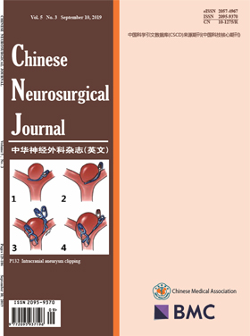
Retrospectively analyzed the clinical characteristics and follow-up data (completely recorded) of 123 patients with 128 aneurysms were treated. 20 cases were treated as control group from October 2013 to December 2013. Since January 2014, aneurysms were classified base on the 20 cases of aneurysm imaging data. 103 patients were treated as experimental group, the classification of aneurysms previously proposed was used to estimate the way of surgery, and the guiding value of the genotype was verified according to the intraoperative findings. The proposed aneurysm classification is based on the virtual surface of the aneurysm and the parent artery, the aneurysm neck was classified as follows: subtype I, the curved surface of the neck is a single curved surface; subtype II, the neck is hyperboloid; subtype III, neck is a three-curved surface. Aneurysms were divided into further subtypes according to the ratio of the width of the aneurysm neck surface and the length of the artery circumference: subtype A, the ratio of the aneurysm neck surface to the parent artery was not more than 0.5; subtype B, more than 0.5. There are some clamping methods include simple, sliding, interlocking and hybrid.
In the control group, patients did not undergo a suitable clipping scheme without classification of aneurysm neck (unclassed clipping). While causing the occurrence of occlusion adverse events, including neck residual, Tumor artery stenosis, electrophysiological changes, the lack of blood supply and so on. The experimental[page1image12073600]group was analyzed by using a predetermined clipping scheme (classed clipping), and the use of aneurysms clamps was approximately the same as expected. Compared the preoperative assessment with the actual situation, the consistency of the control group was 50% and the experimental group was 96%. Adverse events of classed clipping is 2%, another is 60%. There is a significant difference between the two groups (P < 0.05).Classed clipping of subject IA and IB are simple (mean 1.2 and 1.3 clips); classed clipping of subject IIA is simple and interlocking(mean 1.2 clips); classed clipping of subject IIB is sliding and hybrid(mean 2.05 clips); classed clipping of subject IIIA and IIIB are hybrid(mean 2.3 clips).
There is a higher consistency in surgery through the above classification of preoperative assessment of clipping. There was no adverse event of intracranial aneurysm clipping in the clipping mode selected by the above classification, and satisfactory surgical clipping rate was achieved and no recurrence was found.
Surgical interventions including ventriculostomy and ventriculo-peritoneal shunt were usually administrated in pineal germ cell tumor patients with symptomatic hydrocephalus. Considering higher sensitivity of germinoma to anti-tumor therapy, we explored emergency irradiation as non-invasive measure in this situation.
Data of 35 germinoma patients with symptomatic hydrocephalus who received emergency irradiation of 3. 4 Gy/2f were studied retrospectively. The maximum width of frontal horn and the minimum width of trunk of corpus callosum (TCC) were measured to evaluate hydrocephalus changing. Besides, mean deviation (MD) of Humphrey perimetry was employed to evaluate visual field defect. Correlations between hydrocephalus changing and clinical factors, including age, percentage of tumor regression, radiographic re-evaluation interval, and serum beta-human chorionic gonadotropin (β-HCG) level, were analyzed.
The median maximum diameter and volume of pineal lesions was 27 mm (range 10–55 mm) and 6.5cm3 (range 0.4–74.1 cm3), respectively. At median 8 days after irradiation, the median percentage of tumor remission was 55% (range 10–100%). The median maximum width of FN and the median minimum width of TCC were 11.6 mm and 39.0 mm, and 8.0 mm and 31.4 mm, before and after irradiation, respectively. The improvement of both parameters reached significant level (p < 0.001). However, none clinical factor was found to have correlation with their improvement. In 14 patients with paired data of pre- and post-irradiation MD, its change did not reach the significant level for both eyes. All patients successfully received subsequent chemoradiotherapy without surgical intervention.
Emergency irradiation of 3.4 Gy/2f was an effective non-invasive measure to relief hydrocephalus in pineal germinoma patients.
Posterior circulation intracranial aneurysm (IA) treatment remains challenging, due to the anatomy of the area and the high rupture possibility. Endovascular treatment seems to be more suitable for these aneurysms, but studies focused on endovascular treatment demonstrate a high rate of re-intervention needing. A meta-analysis might offer a clearer view, being useful in a more effective treatment planning.
A systematic search was performed, using the PubMed database platform. The final article pool contained 20 articles. Studied parameters were operative mortality, late mortality, permanent neurologic deficit (PND), and the need for re-intervention (Re-int). We divided patients into two subgroups, those with ruptured and those with unruptured aneurysm. Statistical analysis was performed using appropriate software.
In the total population (645 patients), there was a superiority of coiling over clipping in terms of PND and of coiling in terms of Re-int. As regards mortality, there was no clear superiority of one method over the other.
The current study came to the conclusion that there is a superiority of coiling over clipping in terms of PND. On the other hand, clipping seems to be superior to coiling in terms of the need for re-intervention. As regards mortality (both operative and late), there is no clear superiority of one method over the other. Studying subgroups of patients (ruptured and unruptured posterior circulation IAs), in terms of PND, there is no superiority of one method over the other. The same goes for Op-Mo on ruptured aneurysms.
Various computed tomography (CT) appearances of cerebral venous sinus thrombosis (CVST) were associated with different prognosis and the patients with large intracranial hematoma will have adverse outcomes, but no in-depth study of non-contrast CT image appearances was carried out. We aimed to test the hypothesis that non-contrast CT image characteristics on admission are associated with and predict the outcome of CVST at 3 months.
Three hundred and six patients with CVST between 2008 and 2017 were collected. Age, sex, onset of CVST(acute, subacute, or chronic), etiology, clinical manifestations, midline shift, occluded venous sinus, location of infarction, non-contrast CT image characteristics, and the 3-month outcome were recorded. In addition, we established a non-contrast CT image-based classification and grading system to test the hypothesis; the CVST patients were classified into four grades (namely non-contrast CT image-based classification): grade I, no obvious abnormality; grade II, simple vein infarction without hemorrhage or with subarachnoid hemorrhage; grade III, cerebral venous infarction with subarachnoid hemorrhage; and grade IV, cerebral vein infarction with hematoma. All enrolled patients had received subcutaneous injections of low molecular weight heparin subcutaneous injection for 14 days. Thereafter, oral anticoagulant therapy with warfarin was continued. Patients with epilepsy were given antiepileptic drugs, and patients with cerebral herniation received decompressive craniotomy.
Our observational findings revealed that midline shift (> 5 mm), location of lesion (frontal lobe and temporal lobe), and cerebral venous infarction with subarachnoid or hematoma (grade III and IV) were associated with 3-month poor outcome (p < 0.05); the respective increased risks were 12.730 [risk ratio (RR) 12.730, 95% confidence interval (CI) 1.680–96.490, p = 0.014], 46.538 (RR 146.538, 95% CI 6.222–348.079, p = 0.000), 32.549 (RR 32.549, 95% CI 2.180–486.104, p = 0.012), 37.725 (RR 37.725, 95% CI 2.051–693.778, p = 0.015), and 93.164-fold (RR 93.164, 95% CI 11.137–779.328, p = 0.000). However, seizure, hemiplegia, location of occluded venous sinus (super sagittal sinus and deep venous systems), location of infarction (parietal lobe), and non-contrast CT image-based classification (I) were not correlated with the adverse outcome (p > 0.05).
Our findings suggested that non-contrast CT image characteristics on admission were associated with and predict the 3-month outcome of CVST. However, the ultimate conclusions need to be confirmed by a large sample of CVST patients at multiple institutions.
The accuracy and safety of pedicle screw insertion was markedly improved with the introduction of intraoperative three-dimensional navigation system during the last decade. This study aimed to evaluate the accuracy of pedicle screw placement using O-arm-based navigation system versus conventional freehand technique.
We reviewed the accuracy of 341 thoracic (n = 173) and lumbosacral (n = 168) pedicle screws placed in 60 consecutive patients using either O-arm-based navigation or freehand technique in the Department of Neurosurgery of Beijing Tsinghua Changgung Hospital between January 2015 and June 2018. Patient-specific characteristics, treatment-related characteristics, and screw-specific accuracy were analyzed. The accuracy of pedicle screw placement was measured by Gertzbein-Robbins scale and screw grades A and B were clinically acceptable.
One hundred ninety-one screws were inserted in the O-arm-based navigation group and 150 in the freehand group. One hundred eighty-three (95.81%) clinically acceptable screws were placed in the navigation group and 135 (90.00%) in the freehand group (p = 0.034). Twenty-three (6.74%) screw revisions were performed in the two groups (8 screws in the navigation group and 15 screws in the freehand group) and significant difference was observed in thoracic spine (p = 0.018), while no statistical significance was presented in lumbosacral spine (p > 0.05). Twenty-four (12.57%) screws in the navigation group and 24 (16.00%) in the freehand group violated the cortex (p > 0.05). Medial screw deviation was the most common problem in the two groups.
The O-arm-based navigation exhibits higher accuracy for pedicle screw insertion than the freehand insertion technique.
Autophagy is an intracellular degenerative pathway which is responsible for neuronal survival. Under the condition of nutrient deprivation, autophagy can lead to dysfunction in memory consolidation. AMPK/mTOR pathway is currently the most studied autophagy mechanism, while recently researchers have proved ACSS2 can also affect autophagy. ACSS2 is phosphorylated at Ser659 by AMPK and then forms a translocation complex with Importin α5 to translocate into the nucleus. This process interacts with TFEB, resulting in upregulated expression of lysosomal and autophagosomal genes. These upregulations inhibit synaptic plasticity and hence memory functions. On the other hand, ACSS2 is also recognized as a regulator of histone acetylation. After recruiting CBP/p300 and activating CBP’s HAT activity in the nucleus, ACSS2 maintains the level of localized histone acetylation by recapturing acetate from histone deacetylation to reform acetyl-CoA, providing substrates for HAT. The increase of histone acetylation locally enhanced immediate early gene transcription, including Egr2, Fos, Nr2f2, Sgk1, and Arc, to benefit neuronal plasticity and memory in many ways.
Cranial hemophilic pseudotumor (cHPT) is a very rare disease, which is easy to misdiagnose. It is also difficult to manage such patients. We reported the first case of occipital cHPT.
Here, we presented a rare case of an occipital bone mass in a 3-year-old boy who was diagnosed with hemophilia A. The mass was misdiagnosed as an aneurysmal bone cyst by pathological examination. After resection, the patient underwent one-stage cranioplasty. However, the patient was admitted again for hematoma caused by an invasive procedure. A second surgery and one-stage cranioplasty were performed at the same time. A follow-up 3 months after discharging showed the patient was uneventful, and the titanium mesh was well fixed.
The diagnosis of cHPT requires the combining of history, radiological examination, and pathological examination. Resection is the best choice for symptomatic cHPT. Replacement treatment and less invasive treatment can make perioperative management safer. One-stage cranioplasty for resection of an occipital cHPT can improve the quality of life.
Pulmonary alveolar proteinosis (PAP) poses a risk of opportunistic infections with a variety of organisms with Nocardia being the most common pathogen followed by mycobacteria and fungi.
A 7-year-old female child, presented with headache and multiple episodes of vomiting. There was no fever or altered sensorium. On examination, there were no focal deficits or cranial nerve palsies. An MRI brain showed a small T2 hyperintense lesion in the left superior parietal lobe suggestive of an abscess. She was diagnosed as PAP based on CT chest and bronchioloalveolar lavage 7 months earlier and treated with corticosteroids. A left parieto-occipital craniotomy was done with drainage of abscess and abscess wall excision. Histopathology revealed a suppurative lesion with slender septate acute angle branching hyphae which were positive on fungal stains. Culture done on the pus was positive for Aspergillus fumigatus. The patient was treated with voriconazole and stable at 1 year follow-up.
Opportunistic infections are common in patients diagnosed with PAP. High index of clinical suspicion and early diagnosis are important for favorable outcome.
CURRENT ISSUE