MedNexus
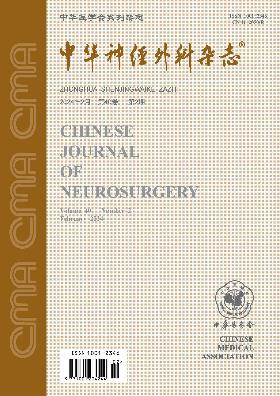
Volume 40 · Issue 02 · 2024
MedNexus
Surgical craniotomy has certain limitations in the treatment of drug-refractory epilepsy. In recent years, new minimally invasive surgical techniques such as stereoencephalogram-guided radiofrequency thermocoagulation and magnetic resonance-guided laser interstitial hyperthermia have been developing continuously, and have become an important treatment choice for drug-refractory epilepsy. This article focuses on the advantages of new minimally invasive techniques, appropriate selection of surgical indications, standardized application and popularization.
To explore the surgical method and clinical efficacy of MRI-guided laser interstitial thermal therapy (MRgLITT) to destroy epileptogenic lesions in the deep brain.
A retrospective analysis was conducted on the clinical data of patients with drug-refractory epilepsy who received MRgLITT at the Neurosurgery Center, Beijing Tiantan Hospital, Capital Medical University from August 2020 to December 2022. In those patients, the epileptogenic zone was located deep in the Sylvian fissure or next to the midline. Patients underwent preoperative multidisciplinary assessment to locate the epileptogenic zone, and a surgical plan was designed based on the assessment results. Postoperative outpatient or video phone follow-up was used to evaluate the patient′s postoperative seizure free rate and complications.
A total of 15 patients with drug-refractory epilepsy were included. The epileptogenic zone was located in the insula in 4 cases and next to the midline area in 11 cases. For insular lesions, transfrontal or transparietal approaches were often used to puncture parallel to the direction of the insular sulcus. Epileptogenic lesions in the midline area were often punctured via transfrontal or transparietal approaches, nearly parallel to the midline. In cases of focal cortical dysplasia, the main purpose was to ablate the bottom of the sulcus. The success rate of MRgLITT was 15/15. All patients were followed up, and the follow-up time was 18.2±5.4 months (12 to 36 months). By the last follow-up visit, the seizure free rate was 11/15, and only 1 case had long-term foot dorsiflexion disorder.
For patients with drug-refractory epilepsy whose epileptogenic zone was located deep in the brain, MRgLITT can help accurately locate the epileptogenic zone and clarify the scope of ablation, thereby achieving ideal surgical efficacy with few and mild complications.
To observe the disease characteristics and surgical efficacy in children with drug-resistant epilepsy caused by GATOR1 complex mutation-associated malformations of cortical development (MCD).
A retrospective analysis was conducted on the clinical data of 35 children with GATOR1 complex mutation-associated MCD who had drug-resistant epilepsy and underwent surgical treatment at the Pediatric Epilepsy Center of Peking University First Hospital from January 2015 to December 2020. The genetic characteristics, clinical features, surgical methods and efficacy were summarized and analyzed. Postoperative follow-up examinations were conducted at 3, 6, 12 months post operation and annually thereafter. Surgical efficacy was assessed based on the outcome of seizure control (Engel class Ⅰ). The postoperative seizure control curves of the patients were plotted using the Kaplan-Meier method, and the surgical efficacy of patients with different gene mutations was analyzed using the log-rank test.
Among the 35 children, 25 (71.4%) had DEPDC5 mutations, 4 (11.4%) had NPRL2 mutations, and 6 (17.2%) had NPRL3 mutations. The most common affected lobe was the frontal lobe (23 cases, 65.7%), while involvement of the occipital lobe was the least common (9 cases, 25.7%). Twenty-eight (80%) children underwent radical surgery directly, and 7 (20%) children received stereotactic radiofrequency thermocoagulation (RFTC) and experienced recurrence. Among them, 1 child continued oral medication, and the remaining 6 children underwent radical surgery. Among the 34 children who underwent radical surgery, 21 underwent focal resection, and 13 underwent disconnection surgery. The postoperative pathological diagnosis was MCD, predominantly focal cortical dysplasia (FCD) type Ⅱa (20 cases, 58.8%). The follow-up duration [M(Q1, Q3)] for the 35 children was 32 (21, 44) months. At the last follow-up, among the 34 children who underwent radical surgery, 27 (79.4%) achieved seizure control. One child treated with RFTC and medication only achieved Engel Class Ⅳ. At the last follow-up, the seizure control rates for children with DEPDC5, NPRL2, and NPRL3 mutations were 22/25, 3/4, and 2/6, respectively. The log-rank test showed that the surgical efficacy of children with DEPDC5 mutations was superior to those with NPRL2 and NPRL3 mutations (P=0.006).
Observations indicate that among children with drug-resistant epilepsy caused by GATOR1 complex mutation-associated MCD who undergo surgical treatment, there is a higher prevalence of DEPDC5 mutations. The lesions mainly involve the frontal lobe, and the most common pathological change is FCD type Ⅱa. Epilepsy surgery is an effective treatment method, with better efficacy observed in patients with DEPDC5 mutations compared to those with NPRL2 and NPRL3 mutations.
To explore the physiological function of the amygdala and its role in the localization of epileptogenic zone (EZ) by analyzing the symptoms produced by electrical stimulation of the amygdala in drug-refractory epilepsy patients.
A retrospective analysis was conducted on the electrical stimulation data of 49 drug-refractory epilepsy patients who underwent stereoelectroencephalography (SEEG) electrode implantation at the Department of Functional Neurosurgery, Xuanwu Hospital, Capital Medical University from January 2017 to April 2023. According to the subject report and clinical observation of the clinical symptoms produced by the electrical stimulation of the amygdala, statistical analysis was conducted on the EZ of epilepsy patients in whom electrical stimulation induced the aura of seizures.
Among 49 patients, a total of 55 SEEG electrodes were implanted in the amygdala, with 192 contacts located in the amygdala. After electrostatic stimuli, 44 patients developed a total of 174 clinical symptoms, including 40 (23.0%) cases of emotional reactions, 52 (29.9%) cases of vegetative symptoms, 33 (19.0%) cases of somatic sensations, 32 (18.4%) cases of head sensations, 10 (5.7%) cases of visual symptoms, 3 (1.7%) cases of olfactory symptoms and 4 (2.3%) cases of motion symptoms. Of the 174 clinical symptoms of 44 patients, the clinical symptoms of 82 (47.1%) procedures of stimulations in 22 (50.0%) patients were auras. The EZ was located in the amygdala and hippocampus in 39 procedures (47.6%, 9 patients), the amygdala in 9 procedures (11.0%, 1 patient), the hippocampus in 17 procedures (20.7%, 7 patients), the frontal lobe in 13 procedures (15.9%, 3 patients) and the insula lobe in 4 procedures (4.9%, 2 patients).
Electrical stimulation of the amygdala in drug-refractory epilepsy patients can induce a variety of functional reactions. Some may be the reaction of the amygdala itself, and some may be the aura of epileptic seizures. It suggests that the amygdala participates in a number of physiological functions of the body and play a role in key nodes of the seizure network.
To explore the safety and effectiveness of vagus nerve stimulation (VNS) with low cervical single incision in the treatment of drug-refractory epilepsy (DRE).
The clinical data of 78 DRE patients admitted to the Epilepsy Center of Qilu Hospital of Shandong University from June 2016 to May 2022 were retrospectively analyzed. Among them, 57 cases underwent VNS through a low cervical single incision (single-incision VNS group), and 21 cases underwent VNS using traditional double incisions (double-incision VNS group). Postoperative outpatient or telephone follow-up was used to evaluate the surgical efficacy based on the control of epileptic seizures (including responder rate and seizure-free rate). The baseline characteristics, operation time, intraoperative blood loss, surgical efficacy and occurrence of postoperative complications were compared between the two groups of patients.
There were no statistically significant differences between the single-incision VNS group and the double-incision VNS group in terms of age, gender, disease duration, epileptic seizure type, operation duration, or intraoperative blood loss (all P>0.05). Comparing the two groups of patients, there were no statistically significant differences in the responder rate (χ2=3.00) or seizure-free rate (χ2=2.00) at 6, 12, or 24 months after surgery (all P>0.05). There was no statistically significant difference in the incidence of postoperative infection or poor healing of incision between the two groups (P>0.05), but the incidence of transient hoarseness [1.8%(1/57) vs. 14.3%(3/21)]or cough [3.5%(2/57) vs. 19.0%(4/21)] was lower in the single-incision VNS group than in the double-incision group (both P<0.05).
The surgical efficacy of single-incision VNS and double-incision VNS in the treatment of DRE are similar, but the former has a lower incidence of postoperative complications and can be used as an alternative VNS surgical method.
To explore the application value of stereoelectroencephalogram (SEEG) electrode implantation in the preoperative evaluation of refractory focal epilepsy.
A retrospective analysis was performed on the clinical data of 24 patients with refractory focal epilepsy who were treated in the Neurosurgery Center, No.988 Hospital of the PLA Joint Logistics Support Force from January 2018 to December 2022 using a relatively small number of SEEG electrodes. Based on the patient′s symptoms, ictal and interictal video-EEG, brain CT, MRI, positron emission tomography (PET)-CT and other imaging data as well as anatomo-electrophysiology-clinical correlation, the hypothesis regarding the seizure onset and propagation network was made. SEEG electrodes were implanted into relevant targets of the seizure network. SEEG monitoring was performed to determine the seizure onset zone. Electrical cortical stimulation (ECS) was administered to induce habitual seizures to verify the epileptogenic zone and locate functional areas. Radiofrequency thermal coagulation (RFTC) was then performed. Those who still had epileptic seizures underwent secondary craniotomy to remove the epileptogenic zone. The Engel classification system was used to evaluate the effect of seizure control.
A total of 126 electrodes were implanted in 24 patients, with 3 to 8 electrodes implanted in each case, with an average of 5.3±1.1 electrodes/case. There were 6 cases of unilateral electrode implantation and 18 cases of bilateral electrode implantation. The error between the actual target point and the planned target point was 1.61±0.88 mm in 24 patients. No patient had additional implanted electrodes. The SEEG monitoring time was 10.1±2.6 days, and 3 to 20 habitual seizures were recorded. ECS induced habitual seizures in 15 cases, non-habitual seizures in 5 cases, and no seizures in 4 cases. The epileptogenic zone was identified in 23 cases, including 10 cases in the temporal lobe, 10 cases in the frontal lobe, and 3 cases in the occipital lobe. The seizure onset could not be located in 1 case. Twenty-two cases were treated with RFTC. Post RFTC, there were 9 cases developing headache, 1 case of contralateral upper limb weakness caused by severe cerebral edema, and 1 case of asymptomatic subdural hematoma. There were no complications such as intracranial infection, cerebrospinal fluid leakage, electrode breakage or displacement. Two cases were not treated with RFTC, of which the hypothesis could not be verified in one case and the epileptogenic area was overlapped with the motor and speech areas in one case. Both cases improved after adjusting anti-seizure medications. All 24 patients received clinical follow-up, and the follow-up time [M(Q1, Q3)] was 12.5(10.2, 26.8) months. The last follow-up showed that among the 22 patients who underwent RFTC treatment, 1 case was Engel grade Ⅰ and 21 cases were grade Ⅱ, and 16 of underwent epileptogenic zonectomy at the second stage. After the resection, the epileptic seizures disappeared in 13 cases (Engel grade Ⅰ), 3 cases had no improvement (Engel grade Ⅱ), and 5 cases were planned to undergo secondary electrode implantation and RFTC treatment.
With reasonable grasp of indications, implantation of a relatively small number of SEEG electrodes can help determine the location of the epileptogenic zone, guide surgical resection of the epileptogenic zone, the postoperative effect is good, and has certain application value in the preoperative evaluation of refractory focal epilepsy.
To explore the initial efficacy of the domestic closed-loop self-responsive neurostimulation system in the treatment of drug-resistant epilepsy.
Six patients with refractory epilepsy admitted to the Neurosurgery Department of the Second Affiliated Hospital of Zhejiang University School of Medicine from March 2022 to November 2022 were prospectively enrolled and treated with a closed-loop self-responsive neurostimulation system. The effectiveness and safety of the treatment were evaluated in the outpatient clinic 4, 7, 10, and 13 months after the start of the stimulation, including the seizure control effect, improvement in quality of life, and occurrence of adverse reactions. The seizure control effect was assessed based on the frequency, duration, and number of days with the seizures in a month.
The operations of all 6 patients were successfully completed. Among them, 4 patients were implanted with deep electrodes (2 per case) in the bilateral hippocampi, 1 patient was implanted with 2 cortical electrodes in the left central cortex, and 1 patient was implanted with 1 deep electrode and 1 cortical electrodes in the left hippocampus + left temporal lobe respectively. Stimulation parameters (median) included the amplitude of 4.0 mA, frequency of 200 Hz, pulse width of 160 μs and duration of 100 ms. Five cases completed the 13-month follow-up after startup, and one case only completed the 11-month follow-up. At 10 months and 13 months follow-up after the device was turned on, the patient′s seizure frequency decreased[M (Q1, Q3)] by 39% (10%, 46%) and 44% (21%, 49%) respectively compared with the preoperative baseline. By the last follow-up, the duration of epileptic seizures was reduced by ≤10% in 4 patients and by >30% in 2 patients; the number of seizure days was reduced by ≤10% in 4 patients and by >30% in 2 patients. Compared with the preoperative baseline, there were no statistically significant differences in the patients′ Hamilton Depression Rating Scale, Quality of Life Scale 31, Hamilton Anxiety Rating Scale or Montreal Cognitive Assessment scores 7 months after the start of stimulation (all P>0.05). As of the last follow-up, no surgery- or device-related adverse events occurred in any patient.
Preliminary observations indicate that treatment with a domestic closed-loop self-responsive neurostimulation system can reduce the frequency of epileptic seizures in patients with refractory epilepsy to a certain extent, and no related adverse events have been observed.
To compare the diagnostic accuracy and diagnostic advantages of prenatal ultrasound and fetal MRI for different types of fetal spinal neural tube defects (SNTDs), and to summarize the characteristic manifestations of prenatal ultrasound and fetal MRI for various types of SNTDs.
We prospectively followed up 161 fetuses admitted to the Neurosurgery Clinic of the First Medical Center of Chinese PLA General Hospital from July 2020 to February 2023 who were considered to have SNTDs in prenatal diagnosis and confirmed by spine MRI examination after birth. A retrospective review of their prenatal and postnatal imaging data was performed. Postnatal MRI examination results were taken as the diagnostic gold standard. The McNemar test was used to compare the diagnostic accuracy of prenatal ultrasound and fetal MRI for various types of fetal SNTDs.
A total of 14 fetal SNTDs were included in the study. The diagnostic accuracy of prenatal ultrasound for spina bifida (P=0.001), intraspinal cyst (P=0.002) and dermal sinus (P=0.002) was higher than that of fetal MRI, while for intraspinal lipoma (P<0.001) and lipomingocele (P=0.039), the diagnostic accuracy of prenatal ultrasound was lower than that of fetal MRI. Prenatal ultrasound was more accurate in diagnosing segmental spinal dysgenesis than fetal MRI (18/18 vs. 7/18); fetal MRI was more accurate than prenatal ultrasound in diagnosing lipomyomeningocele (4/4 vs. 3/4), sacrococcygeal hypoplasia (2/3 vs. 0/3), syringomyelia (2/4 vs. 0/4), caudal regression syndrome (1/1 vs. 0/1), myelomeningocele (6/6 vs. 3/6); for meningocele, tight filum terminale and sacrococcygeal teratoma, the diagnostic accuracy of prenatal ultrasound was the same as that of fetal MRI. For each type of fetal SNTDs, the manifestations of prenatal ultrasound and fetal MRI had their own characteristics.
Prenatal ultrasound and fetal MRI have different advantages in diagnosing fetal SNTDs. Fully understanding the characteristic prenatal imaging manifestations of SNTDs can improve the accuracy of prenatal diagnosis.
To investigate the predictive value of intraoperative monitoring of facial nerve motor evoked potentials (FNMEPs) on facial nerve function (FNF) post vestibular schwannoma (VS) surgery.
A retrospective analysis was conducted on the clinical data of 60 patients with VS who were admitted to the Neurosurgery Center, Beijing Tiantan Hospital, Capital Medical University from May 2020 to December 2020. FNMEP was recorded on the orbicularis oculi, orbicularis oris and mentalis muscles of all patients during the operation, and the FNMEP amplitude decrease rate when the dura mater was sutured was determined. The patients′ facial nerve function was evaluated using the House-Brackmann facial nerve function grading system (referred to as H-B classification) 1 week and 6 months after surgery, and they were divided into a group with good facial nerve function (H-B classification ≤ grade Ⅱ) and a group with poor facial nerve function (H-B classification > grade Ⅱ). The clinical data of the two groups of patients were compared, and indicators with P < 0.20 were included in multivariate logistic regression analysis to explore the predictive factors of postoperative facial nerve function outcomes. The receiver operating characteristic (ROC) curve and area under the curve (AUC) were used to determine the critical value and predictive efficacy for predicting facial neurological outcome after VS surgery.
Among 60 patients, the successful induction rates of FNMEPs from the orbicularis oculi, orbicularis oris and mentalis muscles were 40.0% (24/60), 91.7% (55/60) and 98.3% (59/60) respectively. Total tumor resection was achieved in 48 patients (80.0%) during surgery. The proportion of patients with good facial nerve function at 1 week and 6 months after surgery accounted for 26.7% (16/60) and 68.3% (41/60) respectively. The results of univariate analysis showed that 1 week after surgery, the differences in the amplitude reduction rates of FNMEP in the orbicularis oculi and orbicularis oris muscles between the two groups were statistically significant (both P<0.05). Six months after surgery, there were statistically significant differences in the maximum diameter of the tumor and the amplitude reduction rate of FNMEP in the orbicularis oculi, orbicularis oris and mentalis muscles between the two groups (all P<0.05). The results of multivariate logistic regression analysis showed that the amplitude decrease of FNMEP in the orbicularis oris muscle was the predictor factor of facial nerve function outcome at 1 week after surgery (OR=1.03, 95%CI: 1.01-1.06, P=0.015) and 6 months after surgery (OR=1.05, 95%CI: 1.02-1.08, P=0.001), and the critical values were 33.2% and 48.7% respectively (AUCs: 0.736 and 0.829 respectively, both P<0.05).
Intraoperative FNMEP monitoring, particularly its recording results on the orbicularis oris muscle, can effectively reflect the outcome of postoperative facial nerve function in patients with VS. It is recommended that the amplitude decrease rate >50% be used as an intraoperative early warning standard.
To investigate the efficacy and safety of double-stent combined with intermediate catheter aspiration technique as a rescue treatment in the acute cardiogenic intracranial large vessel occlusion.
The clinical data of 20 patients with acute cardiogenic intracranial large vessel occlusion who received rescue treatment with double stents from January 2019 to June 2023 in the Department of Neurointervention, Luoyang Central Hospital Affiliated to Zhengzhou University were retrospectively analyzed. Conventional thrombectomy and rescue measures failed more than 3 times in all patients before the application of the two-stent combined with intermediate catheter aspiration technique remedy. The recanalization of occluded blood vessels was evaluated immediately after surgery. Modified Thrombolysis in Cerebral Infarction (mTICI) grade ≥2b indicated good recanalization. The modified Rankin Scale (mRS) was used to evaluate neurological deficits 90 days after surgery, with ≤2 points indicating good outcome and 3-6 indicating poor outcome.
Among the 20 patients, 17 patients had good vessel recanalization (mTICI ≥ grade 2b), 1 patient had partial vessel recanalization (mTICI grade 2a), and 2 patients had failed vessel recanalization. Thrombus escape occurred in 1 case, vasospasm in 10 cases, subarachnoid hemorrhage in 3 cases, and symptomatic cerebral hemorrhage in 3 cases. After 90 days of follow-up, 6 cases had a good outcome; 14 cases had a poor outcome, including 10 cases with mRS of 3 to 5 points and 4 cases with mRS of 6 points (death).
As a rescue treatment for acute cardiogenic intracranial large blood vessel occlusion, double stents combined with intermediate catheter aspiration can recanalize most of the blood vessels, and most of the surgery-related complications can be recovered after treatment.
To explore the surgical technique and clinical efficacy of microchannel surgery via the Wiltse intermuscular approach for the treatment of far lateral lumbar disc herniation (FLLDH).
We retrospectively analyzed the clinical data of 7 cases of FLLDH (intervertebral foraminal type Ⅲ) admitted to the Department of Neurosurgery, the First Affiliated Hospital of Shandong First Medical University (Shandong Provincial Qianfoshan Hospital) from September 2021 to June 2023. All those patients had severe pain in the lumbar back and numbness and hypesthesia in the affected lower limbs before surgery. The locations of surgical access and microchannels were determined according to preoperative lumbar CT and MRI scans. The herniated disc was microsurgically resected in the microchannel through the Wiltse intermuscular approach. The patients were followed up by telephone or outpatient clinic at 2 weeks, 1, 3, 6 months and 1 year after surgery. The degree of postoperative pain relief and the presence or absence of new-onset low back pain were assessed based on the visual analogue scale (VAS). The stability of the spine and damage of the small joints were assessed based on lumbar spine CT and three-dimensional reconstruction. The clinical outcomes were evaluated based on the modified MacNab criteria.
Surgeries were all successfully completed in 7 patients, with an operation duration of 62±9 min and intraoperative bleeding volume of 15.0(12.5, 20.0) ml. The severe pain in the low back and the numbness and hypoesthesia symptoms in the affected lower limbs of the 7 patients improved significantly after operation. The VAS score was 2.1±0.9 at 2 weeks post operation, which significantly improved compared with preoperative (7.3 ±1.0, paired t=6.79, P<0.001). The lumbar spine CT and three-dimensional reconstruction at 1 month post operation indicated good protection of the small joints and articular eminences, with little damage to the vertebral plate. No lumbar spine instability or deformity was found. The outcomes of 6 patients assessed based on the modified MacNab criteria were excellent in 5 cases and good in 1 case at 6 months post operation. The median follow-up period of the patients was 13 months (3-24 months). By the latest follow-up, all patients had no recurrence of low back pain, lumbar instability or nerve root injury.
Microchannel surgery via the Wiltse intermuscular approach is safe and effective in the treatment of FLLDH. It is associated with good clinical outcomes based on short- to medium-term follow-up.
Hematoma enlargement in hypertensive intracerebral hemorrhage (HICH) is an important factor leading to poor prognosis of patients. This study included 77 patients with HICH admitted to the Department of Neurosurgery of Lhasa People's Hospital from January 2021 to December 2022, and retrospectively analyzed the influencing factors of hematoma enlargement. Hematoma enlargement occurred in 20 (26.0%) of 77 patients. The results of univariate analysis showed that there were no significant differences in age, gender, ethnicity, history of diabetes, history of hyperlipidemia, history of regularly taking antihypertensive drugs, history of smoking and drinking, Glasgow coma score at admission, whether or not they lived at high altitude, modified Rankin scale score at discharge, hemorrhage site, hematoma morphology, hemoglobin, platelet count and D-dimer between patients in the enlarged and unenlarged hematoma group (allP>0.05); Compared with the group with unenlarged hematoma, the proportion of patients with hypertension ≥1 year in the group with enlarged hematoma was higher, the systolic blood pressure at admission was higher, and the initial hematoma volume was larger, and the differences were statistically significant (allP<0.05)。 The results of multivariate logistic regression analysis showed that systolic blood pressure was high at admission (OR=1.05,95%CI:1.02~1.09,P=0.002), large initial hematoma volume (OR=1.09,95%CI:1.02~1.18,P=0.018) is an important risk factor for hematoma enlargement.
To study the spatial structures among the bony structures, muscles, nerves, and arteries in the cranio-orbital region by microdissection, and to provide anatomical basis for neurosurgery in this area.
A total of 8 adult (16 sides) cadaveric head specimens were selected. The cranio-orbital region was exposed layer by layer using a curved incision via the frontotemporal orbito-zygomatic approach. The anatomical structure of the cranio-orbital junction region was observed and the data related to the supraorbital fissure were documented. The bone structures, muscles, nerve courses ophthalmic arteris anatomy and their position relationship were obserred. The diameters of the nerves entering the orbit through the supraorbital fissure were measured as well.
The cranio-orbital junction region was located below the anterior clinoid process. The optic canal connected the cranial fossa and the orbit. The supraorbital fissure connected the orbit to the middle cranial fossa. The lengths of lateral, medial, and superior margins of the supraorbital fissure in the eight cadaveric head specimens were 18.4±6.2 mm, 7.7±1.6 mm, 16.4±5.6 mm respectively. The widths of the supraorbital fissure on the left side and right side were 3.64±0.86 mm and 3.69±0.88 mm respectively. The widths of the left and right supraorbital fissures were 3.64±0.86 mm and 3.69±0.88 mm respectively. The orbital wall consisted of 7 bony structures: frontal bone, zygomatic bone, ethmoid bone, lacrimal bone, pterygoid bone, palatine bone and maxillary bone. The orbit had a roof, a floor, a medial wall and a lateral wall. Except for the medial wall, the other walls had symmetrically arranged bony structures. The extraocular muscles included 7 muscles: levator palpebrae, rectus superioris, rectus inferioris, rectus medialis, rectus externus, obliquus superioris and obliquus inferioris. The intraorbital nerves mainly consisted of 7 nerves: the optic nerve, the trochlear nerve, three branches of the ophthalmic nerve (frontal, lacrimal and nasociliary nerves), the abducens nerve and the oculomotor nerve (upper and lower branches). Except the optic nerve passing through the optic canal, other 6 nerves (7 branches in total) entered the orbit through the supraorbital fissure and their diameters were 0.86±0.42 mm, 1.51±0.53 mm, 0.71±0.36 mm, 0.82±0.47 mm, 1.23±0.41 mm, 1.29±0.48 mm and 1.82±0.48 mm respectively. The ophthalmic artery which was located below the optic nerve entered the orbit through the optic canal. A total of 16 ophthalmic arteries were observed in 8 specimens. In 1 case there was a duplicated trunk and the distance between the upper and lower trunks was 11.2 mm. In the remaining 7 cases, there were single-trunk ocular arteries. The intraorbital segment of ophthalmic artery was divided into 3 segments according to the angle and curvature of ophthalmic artery. The spatial correlations of the bony structures, muscles nerve courses, and ophthalmic arteries were basically constant.
The anatomy of the cranioorbital region is complicated and its space is narrow. Proficiency in the microanatomy and spatial correlations of the region would help physicians protect important anatomical structures during surgery and particularly lower duplicate ophthalmic artery.
To screen candidate genes associated with radiation resistance by large-sample RNA-sequence and cell experiments; to explore the role of Zinc Finger And BTB Domain Containing Protein 7A (ZBTB7A) in radioresistance of glioblastoma (GBM).
A total of 996 gliomas samples with clinical information and RNA sequencing data were extracted from Chinese Glioma Genome Atlas (CGGA): (1) We compared the data of RNA-seq between 226 primary GBM and 134 recurrent GBM, and 15 paired primary and recurrent GBM to validate the relationship between ZBTB7A and radiation resistance; (2) We compared the ZBTB7A mRNA expression between IDH mutant gliomas and IDH wild-type gliomas; (3) Survival analysis was used to explore the association of ZBTB7A mRNA expression with patients survival (148 low-expression samples vs. 148 high-expression samples ). Patients with primary GBM and recurrent GBM who underwent surgery at Beijing Tiantan Hospital Affiliated to Capital Medical University from April 2019 to December 2022 and were pathologically diagnosed (12 cases each) were included. The protein expression of ZBTB7A was detected in those samples by immunohistochemistry. The GBM cell lines (U87, U251 and LN229) received radiation by high-energy X-ray(single exposure: 13 Gy, 6.5 min). The mRNA expression of ZBTB7A were detected by quantitative PCR in 4 h and 8 h after high-energy X-ray exposure. The protein expression of ZBTB7A in U87 and LN229 cell lines were detected by Western blot at 1 h, 2 h, 4 h, 8 h and 12 h after high-energy X-ray exposure (single exposure: 5 Gy, 2.5 min). U87 cells were infected by ZBTB7A shRNA lentivirus and then received radiation by high-energy X-ray (single exposure: 5 Gy, 2.5 min). The DNA damage were detected by comet assay at 1 h after high-energy X-ray exposure. Colony formation assay confirmed the association of ZBTB7A with radiation resistance in U87 cells after high-energy X-ray exposure.
The crossover analysis based on the RNA sequencing data of unpaired and paired samples showed that ZBTB7A was significant up-regulated in recurrent GBM (both P<0.05). The mRNA expression of ZBTB7A was higher in IDH wild-type gliomas than IDH mutant gliomas (P<0.05). Patients with high expression of ZBTB7A had a shorter overall survival (P<0.05) and progression-free survival (P<0.05) than patients with low expression. Immunohistochemistry showed that the protein of ZBTB7A was higher in recurrent GBM than primary GBM (P<0.05). Comparing with U87 and LN229 cells without high-energy X-ray exposure, the mRNA and protein expressions were significantly increased in cells with high-energy X-ray exposure. The protein of H2AX was also increased at 1 h after high-energy X-ray exposure, but it began to decrease at 4 h after high-energy X-ray exposure. ZBTB7A was knocked down in U87 cells by shRNA lentivirus. The DNA damage was significantly increased in cell with high-energy X-ray exposure compared with control cells (P<0.05). The proliferative activity of U87 cells with high-energy X-ray exposure was also lower than control cells (P<0.05).
The mRNA and protein of ZBTB7A are significantly increased in recurrent GBM. High expression of ZBTB7A is associated with shorter survival. ZBTB7A protein could be induced by radiation and might contribute to the radioresistance of GBM.
To investigate the molecular mechanism of BICD1 gene promoting malignant progression of brain glioma based on spatial transcriptomic, single-cell sequencing data and RNA transcriptomic.
Public dataset of spatial transcriptome was employed to analyze the spatial distribution characteristics of BICD1 in normal brain tissues (sample: "248_C" ) and glioblastoma tissues (sample: "275_T" ). Single-cell sequencing data (from 6 148 cells of 14 patients) in Chinese Glioma Genome Atlas (CGGA) database were adopted to decipher the expression difference of BICD1 in different glioma cell types by R-language software. The biological functions of BICD1-related genes were analyzed based on RNA sequencing data in CGGA database (n=693) via the Kyoto Encyclopedia of Genes and Genomes (KEGG) and the Gene Ontology (GO) analyses.
BICD1 was expressed in a wide range in normal brain tissues and specifically expressed at the boundary between glioma stem cells and tumor cells in glioblastoma tissues. Cell subtyping analyses based on single-cell sequencing data showed that BICD1 was highly expressed in astrocytes. In the Neftel subtyping system, cells with BICD1 highly expressed were mainly distributed in astrocytes-like cells and neural progenitor-like cells clusters. KEGG pathway analyses of BICD1-related differential genes showed that high expression of BICD1 was associated with enrichments of tumor-related biological functions such as cellular senescence (P=0.003), proteoglycans in cancer (P=0.004), and enrichments of intracellular transport processes such as actin cytoskeleton regulation(P=0.010). Low expression of BICD1 was associated with enrichments of ribosome-related functions (P<0.001) and biological processes such as oxidative phosphorylation (P<0.001). GO analyses of BICD1-related differential genes showed that cytoplasmic translation (P<0.001), ribosome small subunit assembly (P=0.001), regulation of chromosome separation (P=0.004) and chromosome separation (P=0.004) were significantly correlated with BICD1 expression levels. The expression level of BICD1 was significantly correlated with biological processes such as polyubiquitin modified-dependent protein binding(P=0.046), ubiquitin-ubiquitin ligase activity (P=0.033), negative regulation of protein depolymerization (P=0.010), monoubiquitinated protein deubiquitination (P=0.026), actin filament capping (P=0.007) and actin filament depolymerization (P=0.013).
BICD1 might be involved in protein ubiquitination degradation and cytoskeleton-related intracellular transport in precursor glioma cells, and plays a critical role in the malignant transformation of those cells by regulating biological processes such as cell senescence and cell division.
Calcification is one of the common features of meningiomas, but extensive calcification of tumors is rare clinically. In November 2022, the Neurosurgery Center of Beijing Tiantan Hospital affiliated to Capital Medical University treated a patient with sellar meningioma with extensive calcification by neuroendoscopic expanded transnasal approach. Subtotal tumor resection was performed, and no related complications occurred after operation. The patient recovered well after 7 months of follow-up.
There is no effective treatment for motor dysfunction left over from stroke. Basic experimental studies have shown that deep brain electrical stimulation (DBS) is an ideal therapeutic method with potential; Several clinical studies have also found that patients have improved symptoms after receiving DBS treatment. This article reviews the research progress of DBS in the treatment of motor dysfunction after stroke.
The formation and rupture of intracranial aneurysms are the risk factors that seriously endanger human health and reduce the quality of life. According to the latest statistics, the prevalence rate of intracranial aneurysms in the world is about 7%, and its incidence and mortality rate remain high in China. The abnormal elevation of plasma homocysteine (Hcy) has been confirmed to be closely related to intracranial vascular diseases. Recent studies have shown that it may also be related to the formation and rupture of intracranial aneurysms. The specific mechanism and pathogenic pathway are still unclear. This article reviews the risk factors affecting the existence of plasma Hcy instability, the pathogenesis of intracranial aneurysm formation and the increased risk of rupture, so as to effectively prevent the formation of intracranial aneurysm and reduce the risk of rupture.
Glioblastoma is the most malignant and lethal tumor among central nervous system tumors, but its treatment effect is not satisfactory at present. The most important reason is that there is a process called microvascular mimicry in glioblastoma. This biological process ensures sufficient blood and nutrient supply during tumor occurrence and development, and also explains the current mainstream cognition. The reason for the poor effect of targeted therapy against tumor angiogenesis. At present, the widely recognized mechanisms of microvascular mimicry include: epithelial-mesenchymal transition, local hypoxia, related cytokines and various signaling pathways mediate the formation of microvascular mimicry. This article will review the above-mentioned mechanisms of microvascular mimicry formation in glioblastoma, in order to provide help in the clinical treatment of this disease.
Tethered cord syndrome is a series of neurological dysfunction syndromes caused by spinal cord traction, low conus, ischemia and hypoxia of spinal cord, and neurological degeneration due to various reasons. Because the onset site of this disease is spinal conus, the symptoms are similar to those of orthopedics, urology, pediatrics and other related diseases, and it is easy to be misdiagnosed in clinic. For recurrent and occult complex tethered cord syndrome, there is no consensus on the timing and surgical method of operation. This article reviews the surgical treatment of tethered cord syndrome in recent years in order to strengthen the understanding of the diagnosis and treatment of tethered cord syndrome.
CURRENT ISSUE