MedNexus
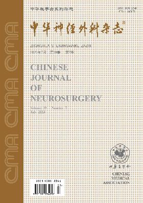
Volume 39 · Issue 07 · 2023
MedNexus
Craniocerebral trauma is still a type of disease that seriously affects the health of people. In recent years, the treatment level of craniocerebral trauma has been continuously improved, but there are still problems such as uneven medical capacity between regions and poor long-term prognostic indicators. The improvement of clinical management quality of craniocerebral trauma becomes the key to further improve its therapeutic effect. This article focuses on the quality control and promotion of craniocerebral trauma treatment in China.
To investigate the clinical features of dural arteriovenous fistula of the hypoglossal canal (HCDAVF) and the efficacy of endovascular embolization treatment.
The clinical data of patients with HCDAVF who underwent endovascular treatment at the Neurosurgical Center of Beijing Tiantan Hospital, Capital Medical University from October 2019 to October 2021 were retrospectively analyzed. Lesions were classified based on the criterion proposed by Björn Spittau. Endovascular embolization via only vein or combined arteriovenous approach was performed depending on the specific situations. All patients were followed up by outpatient clinic visits or telephone regularly. Digital subtraction angiography was used to evaluate the efficacy of endovascular embolization.
A total of 7 patients with HCDAVF were enrolled into this study. The main clinical symptom was pulsatile tinnitus in 3 cases and bulbar conjunctival edema in 4 cases. The fistula was located on the right side of the anterior condylar confluence (ACC) in 4 cases, on the left side of the ACC in 2, and on the right side of the anterior condylar vein and ACC in 1. Successful procedures were performed in all patients. Endovascular embolization was performed only via vein in 6 cases or combined arteriovenous approach in 1 case. Immediate postoperative DSA results showed complete embolization in all 7 cases. No postoperative complications were found. The median time of follow-up was 12 months (range: 6-13 months). DSA results showed that no recurrence was detected in any of the 7 patients and clinical follow-up showed that all clinical symptoms disappeared.
Pulsatile tinnitus and bulbar conjunctival edema are common clinical manifestations in HCDAVF patients. Endovascular embolization of type 1 and type 2 HCDAVFs via only vein or combined arteriovenous approach demonstrates a good safety and efficacy profile.
To preliminarily investigate the safety and efficacy of a novel Kylin flow diverter for the treatment of small intracranial unruptured aneurysms.
We prospectively enrolled 22 patients with small intracranial wide-necked unruptured aneurysms(22 aneurysms in total) who were treated with novel Kylin flow diverter at the Neurovascular Center, the First Affiliated Hospital of Naval Medical University between July 2021 and October 2021. Raymond grading system was used to evaluate the degree of occlusion immediately and 12 months after operation. Clinical and imaging follow-up were performed. At 12 months post procedure, the primary efficacy endpoint was complete occlusion (Raymond grade Ⅰ), and the safety endpoint was newly-onset stroke in the brain area supplied by the treated artery or neurologic death.
All aneurysmal necks were completely covered by Kylin flow diverters(23 in total) and satisfactory adherence between the device and vessel wall was achieved. All patients received 12-month imaging and clinical follow-up. A total of 18 (81.8%) patients met the primary efficacy endpoint. None of them suffered from newly-onset stroke in the brain area supplied by the treated artery or neurologic death.
Preliminary study has shown that treatment of small intracranial unruptured aneurysms with Kylin flow diverter is safe and effective.
To investigate the safety and efficacy of Neuroform Atlas stent-assisted coil embolization for treatment of intracranial aneurysms at middle cerebral artery (MCA) bifurcation.
A retrospective analysis was conducted on the clinical data of 21 patients with aneurysms at MCA bifurcation who underwent endovascular treatment using Neuroform Atlas device in the Department of Neurology, Chinese PLA General Hospital from July 2020 to October 2022. The immediate postoperative embolization degree was assessed using Raymond grading system and perioperative complications were documented. DSA was performed at 6-month follow-up for evaluation of aneurysm embolization degree and related complications.
Among 21 cases of MCA bifurcation aneurysms, single-stent treatment was administered in 18 cases and double-stent treatment in 3 cases. All stents were accurately located and implanted. The successful rate of procedure was 100%. As revealed in immediate angiography after the procedure, there were 3 cases (14.3%) of grade Ⅰ, 1 case (4.8%) of grade Ⅱ and 17 cases (80.9%) of grade Ⅲ. There were 2 perioperative complications documented which included 1 case of in-stent thrombosis developing during surgery and 1 case of postoperative transient ischemic attack. Both of them had no obvious symptom or sign of neurological dysfunction. A total of 13 patients underwent DSA review at 6-month follow-up which revealed grade Ⅰ in 11 cases, grade Ⅱ in 1 case and grade Ⅲ in 1 case. There were 1 case of recurrence, 3 cases of in-stent stenosis of parent vessel and 1 case of blocking of MCA branch on the contralateral side of aneurysm.
It has been preliminarily suggested that application of Neuroform Atlas stent in the treatment of MCA bifurcation aneurysm is associated with high safety and favorable short-term embolization efficacy.
To preliminarily investigate the efficacy and safety of Surpass Streamline flow diverter(FD) in the treatment of intracranial aneurysms.
A retrospective study was performed on the clinical data of 11 patients with intracranial aneurysms treated with Surpass Streamline FD in the Cerebrovascular Division of Interventional Therapy Center, Henan Provincial People′s Hospital from December 2020 to March 2022. There was a total of 14 aneurysms in 11 patients, 13 of which were located in the internal carotid artery and 1 in the V4 segment of the vertebral artery. The whole-brain digital subtraction angiography (DSA) was used to review aneurysm occlusion immediately after surgery and during follow-up and the O′Kelly-Marotta (OKM) grade was used to assess the degree of embolization of aneurysms; The modified Rankin Scale score (mRS) was used to evaluate the neurological function of patients.
A total of 11 surpass streamline FDs were successfully placed to treat 14 aneurysms. Of those, 6 were treated with surpass stream FD only and FD adherence were induced by supplementary stent or Scepter balloon dilation in 5 cases. Perioperative ischemic complications occurred in 1 patient with balloon dilatation and the mRS was 3 points upon discharge. The OKM grade immediately after surgery was grade A in 1 case, grade B in 8 and grade C in 5. All 11 cases underwent clinical follow-up which lasted for 6.0 (5.8, 7.2) months [M(Q1, Q3)]. At the last follow-up, the mRS was 2 points in 1 patient and 0 in the remaining 10 patients. DSA follow-up was obtained in 6 patients, and the median time of follow-up was 6.2 (5.8, 6.4) [M(Q1, Q3)] months. The OKM grade D was reported in 5 cases and grade C in 1. One case developed asymptomatic in-stent stenosis.
Surpass streamline FD shows a high procedural success rate for the treatment of intracranial aneurysms. It is associated with favorable immediate postoperative effect and low complication rate. The short-term follow-up indicates its safety and effectiveness.
To investigate the effectiveness of lantern technique using braided stent (LVIS) in performing coil embolization for ruptured wide-necked posterior communicating artery (PCoA) aneurysms combined with fetal-type posterior cerebral artery (fPCA).
A retrospective analysis was conducted on the clinical data of 24 consecutive patients with ruptured wide-necked PCoA aneurysms combined with fPCA who underwent the lantern technique by using LVIS at the Department of Neurosurgery, Nanjing First Hospital, Nanjing Medical University between January 2015 to January 2020. The embolization outcome was evaluated using the Raymond grading system. The clinical outcome was assessed based on the modified Rankin scale (mRS): 0-2 as favorable outcomes and 3-6 as unfavorable outcomes.
All 24 patients underwent successful aneurysm embolization. There were 2 cases (8.3%) of perioperative transient ischemic attack. Immediate postoperative DSA showed that Raymond grade Ⅰ was achieved in 16 cases, Raymond grade Ⅱ in 4 cases, and Raymond grade Ⅲ in 4 cases. The fPCA was patent in all cases. The average clinical follow-up time of 20 patients was 58.0±9.2 months (25-79 months). The mRS score was 0 in 19 cases and 1 in 1 case. The median imaging follow-up time of 19 patients was 52 months (3-65 months). Raymond grade Ⅰ was achieved in 17 and Raymond grade Ⅱ in 2 cases. Severe in-stent stenosis with PCoA occlusion occurred in 1 case. The PCoA was patent in the remaining patients.
The lantern technique using LVIS stent showed good safety and efficacy in the treatment of ruptured wide-necked PCoA aneurysm combined with fPCA.
To explore the therapeutic effect and clinical experience of microsurgery in the treatment of craniocervical junction dural arteriovenous fistulas (DAVF).
The clinical data of 10 patients with craniocervical junction DAVF treated by microsurgery in the Department of Neurosurgery, the First Affiliated Hospital of Zhengzhou University from May 2020 to April 2022 were retrospectively analyzed. All fistulas were located between the lower edge of the foramen magnum and the cervical 1 vertebral body. The lesion was supplied by radiculo-meningeal artery in 8 cases and by radiculo-meningeal artery and lateral spinal artery in 2 cases. The lesion was drained by vein to the head side in 6 cases and drained to the neck side in 4 cases. Microsurgery was used to clip the fistula or cut off the fistula by electrocoagulation, and intraoperative angiography confirmed the disappearance of the DAVF. Clinical and imaging follow-up was carried out 3 months after operation, and the surgical effect was evaluated by comparing with preoperative clinical symptoms.
All 10 patients achieved imaging cure immediately after surgery with good incision healing, and there was no cerebrospinal fluid leakage. Postoperative symptoms were relieved in 8 cases, aggravated in 1 case, and not improved in 1 case. Two patients developed high fever and headache after operation, and the symptoms disappeared after symptomatic treatment. During the 3-month follow-up, 1 patient′s symptoms improved significantly, and 8 patients′ symptoms disappeared; the other patient′s symptoms did not improve, and DSA showed recurrence of DAVF.
Microsurgical treatment of craniocervical junction DAVF is safe and effective. The key to the success of surgery is to identify the fistula and all the supplying arteries.
To investigate the clinical characteristics, prognosis and prognostic factors of malignant melanomas (MM) in the central nervous system.
A retrospective analysis was conducted on the clinical data of 26 patients with pathologically confirmed central nervous system MM who underwent surgical resection at the Department of Neurosurgery, the First Affiliated Hospital of Zhengzhou University from January 2015 to September 2021. Among the patients, 23 had intracranial MM and 3 had intraspinal MM. Kaplan-Meier method was used to explore the difference of survival between intracranial MM and intraspinal MM. Univariate log-rank test and multivariate Cox regression survival analyses were performed to identify prognostic factors of intracranial MM.
The 26 cases with central nervous system MM had no characteristic clinical manifestations. Preoperative MRI showed short T1 and short T2 signals in some patients, which, however, could not lead to a definite diagnosis. The surgeries were successfully completed in all patients. Total resection was achieved in 16 cases and subtotal resection in 10 cases (including 1 case of intraspinal MM). Postoperative pathological evaluation showed MM in all 26 cases. Detection of the BRAF V600E mutation was carried out in 3 cases and revealed positive in 1 case. All 26 cases were followed up for a median of 9 months (2-58 months). Three patients underwent a second operation after MM recurrence. At the last follow-up, 24 cases died and 2 cases (intracranial MM) survived. Out of the 2 cases, 1 had BRAF V600E mutation and the multiple intracranial metastases almost disappeared at 7 months post targeted therapy. Median survival for the 23 intracranial cases was 8.0 months (4.9-11.1 months) and Kaplan-Meier survival analysis showed that survival rates at 6 months and 1 year post operation were 70% and 24% respectively. The survival periods of 3 intraspinal cases was 49, 15 and 58 months respectively, which were all longer than that of the intracranial cases. Univariate log-rank test analysis for the 23 intracranial cases showed that extent of resection was the only independent prognostic factor for survival (P=0.002), while differences in gender, age, tumor origin (primary or metastatic), tumor location (supratentorial or infratentorial), Ki-67 positive rate (<50% or ≥50%), or postoperative adjuvant therapy were not statistically significant (all P>0.05).
Central nervous system MM is a rare disease with atypical clinical and MRI manifestations and a high misdiagnosis rate. Its established diagnosis should be based on pathological examination. Extent of tumor resection is an independent prognostic factor of intracranial MM. Survival of patients with intracranial MM is shorter than that with intraspinal MM and its overall prognosis remains poor.
To compare the efficacy of conventional face-to-face programming and remote programming after deep brain stimulation (DBS) in patients with Parkinson′s disease (PD).
A prospective study was conducted on 30 PD patients who underwent DBS surgery at the Neurosurgery Center of Beijing Tiantan Hospital, Capital Medical University from April 2019 to December 2021. All patients underwent initial turning-on and first programming at the outpatient clinic one month after surgery, and then programmed once every three months. According to different programming methods, they were divided into remote programming group (15 cases) and conventional outpatient programming group (15 cases). The two groups of patients were evaluated before surgery and 6 months after initial programming. The baseline data before surgery, the score of part Ⅲ of the Movement Disorder Society-Unified Parkinson′s Disease Rating Scale (MDS-UPDRS Ⅲ), the daily levodopa equivalent dose (LEDD), adverse reactions, treatment-related time cost and economic cost, and patient and family satisfaction scores were compared between the two groups.
There was no significant difference in gender, surgical age, disease course, preoperative MDS-UPDRS Ⅲ score, LEDD, and preoperative medication improvement rate between the two groups (all P>0.05), and the two groups were comparable. Six months after initial programming, there was no significant difference in MDS-UPDRS Ⅲ score and motor score improvement rate, incidence of adverse reactions, or LEDD reduction between the remote programming group and the outpatient programming group (all P>0.05). The time spent on programming [M(Q1, Q3)] in the remote programming group was less than that in the outpatient programming group [38 (27, 62) h vs. 120 (72, 216) h, U=25.50, P=0.001] and the cost [M(Q1, Q3)] was less [900 (900, 1 200) yuan vs. 8 000 (1 800, 12 500) yuan, U=11.00, P=0.001]. The overall satisfaction of patients with symptom control at 6 months post initial programming, the satisfaction of family members and carers with the programming process, and the effectiveness of communication with the programming physician were all higher in the remote programming group than in the outpatient programming group (all P<0.05).
Remote programming and outpatient programming can achieve similar therapeutic effects in PD patients after DBS. Remote programming does not increase the incidence of adverse reactions, saves time and economic costs, and has higher patient satisfaction. It is worthy of clinical application and promotion.
To investigate the resolution time of chronic subdural hematoma (CSDH) after middle meningeal artery embolization (MMAE) and its influencing factors.
A retrospective analysis was performed on the clinical data of 41 (53 lesions) CSDH patients treated with MMAE or MMAE combined with burr hole drainage in the Department of Interventional Radiology, the Affiliated Hospital of Yangzhou University from May 2019 to May 2021. The patients′ demographic data, preoperative comorbidities that may affect hematoma absorption or recurrence, whether antithrombotic therapy was administered, preoperative imaging characteristics of hematoma, intraoperative Dyna CT hematoma membrane enhancement grade and postoperative hematoma absorption were documented. For patients with bilateral lesions, hematoma conditions were assessed based on the larger hematoma. Patients were divided into complete absorption group and incomplete absorption group according to whether the hematoma was completely absorbed 4 months after surgery. Univariate and multivariate logistic regression analyses were performed to determine the influencing factors of CSDH absorption.
A total of 53 surgical procedure for CSDH lesions were performed in 41 patients, out of whom 22 patients underwent MMAE combined with burr hole drainage. The maximum diameter of postoperative residual hematoma was 13.5±3.6 mm (10.8-21.5mm). Hematoma recurrence after MMAE was reported in 1 case (2.4%). No MMAE-related complications were observed in this series. All 41 patients were followed up for 17.2±6.1 weeks. The hematoma was completely absorbed in 40 cases (97.6%) at the latest follow-up, in 27 cases (65.9%) at 4-month follow-up and in 39 cases (95.1%) at 6-month follow-up. The median absorption time of CSDH was 14 (12, 20) weeks. Univariate logistic regression analysis showed that compared with complete absorption group (27 cases), the patients were older, proportion of antithrombotic treatment after MMAE was higher, maximum diameter of residual hematoma was larger, proportion of intraoperative Dyna CT hematoma membrane enhancement grade Ⅰ was higher, proportion of patients undergoing MMAE combined with burr hole drainage was lower in the incomplete absorption group (14 cases) (all P<0.05). Multivariate logistic regression analysis showed that the maximum diameter of residual hematoma (OR=0.63, 95%CI: 0.31-0.92, P=0.012), the intraoperative Dyna CT hematoma membrane enhancement grade Ⅰ (OR=0.48, 95%CI: 0.25-0.81, P=0.027) and whether combined burr hole drainage were performed (OR=3.13, 95%CI: 0.37-0.89, P=0.026) were independent factors for complete absorption of hematoma at 4-month follow-up.
After MMAE treatment, CSDH can usually be absorbed within 4 months. Incomplete absorption of CSDH hematoma at 4-month follow-up may be associated with the large maximum diameter of residual hematoma after MMAE, intraoperative CT hematoma membrane enhancement grade Ⅰ and treatment without combined burr hole drainage.
To investigate the occurrence and outcome of epidural fluid (EFC) after cranioplasty (CP) and to explore the risk factors of EFC after CP.
A retrospective study was performed on the clinical data of 201 patients undergoing CP at the Department of Neurosurgery, Shanghai Tenth People′s Hospital from January 2017 to December 2020. The incidence of perioperative EFC was documented. Using univariate and multivariate logistic regression analyses, we studied the effects of age, gender, bone window area, history of hypertension and diabetes, operation opportunity, repair materials, history of in situ repair, cause of craniectomy, bilateral repair, bone window position, preoperative shunt, operation time, combined shunt, duration of wound drainage, first-day drainage flow, total drainage flow, preoperative epileptic seizure, preoperative hydrocephalus, postoperative new hydrocephalus, cerebral hemorrhage, epilepsy and infection on the occurrence of perioperative EFC.
A total of 123 patients (61.2%) developed EFC during the perioperative period, and all recovered after treatment. In the non-EFC group, the bone window was located in the frontal region in 68 cases (87.2%) and in the temporal region in 10 cases (12.8%). In the EFC group, the bone window was located in the frontal region in 117 cases (95.1%) and in the temporal region in 6 cases (4.9%). In the non-EFC group, 15 cases (19.2%) were repaired with polyetheretherketone (PEEK) material, and 63 cases (80.8%) were repaired with titanium mesh. The EFC group had 41 cases (33.3%) repaired with PEEK and 82 cases (66.7%) repaired with titanium mesh. The differences between the two groups were statistically significant (all P<0.05). There was no significant difference in other indicators between the two groups (all P>0.05). Multivariate logistic regression analysis showed that PEEK repair material was an independent risk factor for perioperative EFC (OR=2.05, 95%CI: 1.03 -4.04, P=0.030).
The incidence of EFC after CP seems high and early treatment could lead to a favorable prognosis. PEEK material is a risk factor for EFC after CP.
To explore the structures of the white matter fiber tracts above the external capsule by micro-anatomical study.
Eight formalin-fixed (10%) adult human brains (16 hemispheres) were examined by using Klingler fiber dissection technique with the aid of a microscope at 6-40 magnification. The structures and relationships of the fiber tracts above the external capsule were observed.
There are multiple kinds of fiber tracts above the external capsule. After removing the gray matter, the U fibers were exposed, which were the most superficial and widely distributed fiber tracts. The main fiber tracts between the U fibers and external capsule included the superior longitudinal fasciculus, arcuate fasciculus, middle longitudinal fasciculus, inferior longitudinal fasciculus, extreme capsule, external capsule, claustrum and its fiber tracts. The courses of those fibers tracts and their connections with each other were mainly constant.
The directions and relationships of the fiber tracts that above the external capsule can be confirmed by the method of micro-anatomy. This study can facilitate the formulation of safe and accurate surgical plans by physicians and provide relevant basis for the research of the brain function.
To investigate the biological significance of tumor-associated macrophage (TAM) infiltration in gliomas.
U87, U251 cell lines and human tissue monocytes (THP-1) were used in this study. Human tissue monocytes were induced to differentiate into macrophages and polarized into M2 macrophages. U87 and U251 cell lines (U87+ TAM group, U251+ TAM group) were cultured in M2 macrophage conditioned medium. At the same time, U87 and U251 cell lines were cultured separately as U87 control group and U251 control group. The expression of STAT3, phosphorylated STAT3, JAK2 and phosphorylated JAK2 in each group was detected by Western blot. Furthermore, the effects of macrophages on the proliferation, migration and invasion ability of glioma cells were further confirmed by CCK-8 method, colony formation assay, scratch healing experiment and Transwell chamber experiment.
The results of Western blot showed that the expression of phosphorylated STAT3 (P=0.041) and phosphorylated JAK2 (P=0.020) in the U87+ TAM group and U251+ TAM group was significantly increased compared with the U87 control group and U251 control group. The CCK-8 test showed that compared with the control group, the proliferation rate of cells in the U87+ TAM group and the U251+ TAM group was significantly accelerated (U87: P=0.013 at day 3, P=0.047 at day 5, P=0.016 at day 7; U251: P=0.013 at day 3, P=0.004 at day 5, P=0.007 at day 7). The results of colony formation assay, scratch healing experiment and Transwell chamber experiment showed that compared with the U87 control group and U251 control group, the number of colonies formed, migration ability and invasion ability in U87+ TAM group and U251+ TAM group were enhanced.
TAM infiltration can promote the proliferation, migration and invasion of brain glioma cells.
To investigate the expression and proneural-to-mesenchymal transition (PMT) function of homeodomain-interacting protein kinase-2 (HIPK2) in gliomas.
We first analyzed the expression of HIPK2 in the low-grade gliomas (LGGs) and high-grade gliomas (HGGs) in National Center for Biotechnology Information Gene Expression Omnibus (NCBI-GEO) dataset GSE16011. Based on the GSE16011 dataset, we further analyzed the expression of HIPK2 in proneural subtype, mesenchymal subtype and classical subtype gliomas. According to the HIPK2 expression, the testees were divided into low expression group and high expression group, and Kaplan-Meier survival curve was drawn. Log-rank test was used to compare the survival of glioma patients in the HIPK2 low expression group with that in the HIPK2 high expression group. Human astrocytes (NHA) were set as the control group and Western blot was performed in vitro to assess the expression of HIPK2 in H4, A172, U87 and U251 glioma cell lines. In U87 cells, overexpressed lentiviral vectors carrying the HIPK2 cDNA sequence were used to construct cells overexpressing HIPK2. Construction of control cells was made with lentiviral vectors. H4 cells were transfected with siRNA to knock down the expression of HIPK2. We then studied the effects of HIPK2 on the migration and invasion ability of glioma cells through cell scratch experiment and transwell experiment. We further performed Western blot to detect the effect of HIPK2 on the expression of FN1, CD44 and SNAI2. Finally, Pearson correlation analysis was used to analyze the correlation between HIPK2 expression and FN1, CD44 and SNAI2 expression in GSE16011 dataset.
In the GSE16011 dataset, HIPK2 mRNA expression levels of HGGs was significantly lower than that of LGGs (t=6.86, P<0.001). Lower expression of HIPK2 mRNA was found in mesenchymal subtype and classical subtype gliomas (both P<0.001). Among glioma patients or HGGs, the overall survival of the HIPK2 low expression group was shorter than that of the high expression group (40.8±3.9 months vs. 24.9±3.2 months, P<0.001). In further in vitro experiments, Western blot results showed that the protein level of HIPK2 in H4, A172, U87 and U251 cells was decreased compared with that of NHA group (all P<0.05). The protein level of HIPK2 in U87 overexpression group was upregulated compared with that of the control group (t=24.88, P<0.05). The protein level of HIPK2 in the H4 knockdown group was downregulated compared with the control group (t=26.72, P<0.001). The cell scratch experiments results showed that the cell scratch repair rate of U87 overexpression group was lower than that of U87 control group (18.1%±2.0% vs. 45.6%±4.2%, t=10.23, P<0.001). On the contrary, the cell scratch repair rate of H4 knockdown group was higher than that of H4 control group (60.5%±5.4% vs. 37.9%±4.0%, t=5.83, P=0.004). Transwell experiments results showed that the number of membrane-penetrating cells in the U87 overexpression group was lower than that of U87 control group (92.0±11.9 vs. 193.1±16.2, t=8.75, P<0.001). On the contrary, the number of membrane-penetrating cells in the H4 knockdown group was higher than that of H4 control group (204.0±16.9 vs. 135.1±15.0, t=5.27, P=0.006). Western blot results showed that the protein levels of FN1, CD44 and SNAI2 in the U87 overexpression group were lower than those of U87 control group (all P<0.001). On the contrary, the protein levels of FN1, CD44 and SNAI2 in the H4 knockdown group were higher than those of H4 control group (all P<0.001). Pearson correlation analysis results showed that the expression of FN1, CD44 and SNAI2 was negatively correlated with HIPK2 expression in GSE16011 dataset (all P<0.05).
HIPK2 is lowly expressed in HGGs and mesenchymal subtype gliomas. Low expression of HIPK2 may promote glioma cell migration and invasion via enhancing PMT process.
Clinically, pituitary metastasis of malignant tumors is common, but metastasis to pituitary stem is relatively rare. At present, the number of cases reported at home and abroad is small. In April 2022, the Department of Neurosurgery of Chengdu Second People's Hospital admitted a patient with pituitary stem metastasis of lung cancer with increased prolactin level as the main manifestation, and underwent craniotomy to remove the lesion. Postoperatively, the patient's electrolyte disorder was corrected and hormone levels returned to normal.
Vertebrobasilar lengthy dilatation is a rare intracranial dissecting aneurysm, and the acute occlusion of the basilar artery caused by it is more rare in clinic. In February 2022, the Department of Neurosurgery of the Second Hospital of Hebei Medical University treated a patient with acute occlusion of the basilar artery caused by lengthy dilatation of the vertebral-basilar artery. He underwent global cerebral angiography + left vertebral-basilar artery stenting for emergency, and was given antiplatelet therapy after operation. On the third day after operation, the patient's consciousness returned to normal, and the muscle strength of both upper limbs was grade IV, and the muscle strength of both lower limbs was grade Ⅲ. The muscle strength of the patients returned to normal (all grade V) at the 2-month follow-up, the modified Rankin scale score was 0, and no new infarction was found on MRI. For such patients, it is safe and effective to use large-size stents to open occluded blood vessels.
Aneurysmal bone cyst is a benign cystic bone tumor. In October 2021, a patient with frontotemporal sphenoid aneurysmal bone cyst was treated by surgical resection combined with titanium plate repair in the Department of Neurosurgery of Lishui People's Hospital. The aneurysmal bone cyst was confirmed by pathological examination. The patient recovered well after surgery; Follow-up until 8 months postoperatively showed no recurrence.
The right aortic arch is a relatively rare variant, usually associated with supraarchal vascular abnormalities. In December 2020, the Department of Neurosurgery of the Second Hospital of Hebei Medical University treated a patient with right aortic arch combined with stenosis of left vertebral artery V2 segment and severe stenosis of right internal carotid artery extracranial segment. The patient was treated with stenting of left vertebral artery V2 segment and endarterectomy of right internal carotid artery. The patient's symptoms were significantly improved without related complications.
Pituitary neuroendocrine tumor is a common intracranial tumor. Some pituitary neuroendocrine tumors show invasive behavior, which can involve sphenoid sinus, cavernous sinus, suprasellar sinus, and even surrounding tissues such as dura mater and slope of skull base, resulting in low total resection rate and high recurrence rate. More effective treatment measures are urgently needed. Immune microenvironment is the local environment on which tumor cells depend. The study of immune microenvironment is helpful to understand the mechanism of occurrence, development and invasion of pituitary neuroendocrine tumors, and discover new immunotherapy targets. This article reviews the related studies of immune microenvironment promoting the aggressiveness of pituitary neuroendocrine tumors.
Traumatic optic nerve injury is a serious complication of craniocerebral trauma that is common in young men. Patients after injury often have visual impairment, and their prognosis is poor. Poor prognostic factors may include absence of light perception after injury, delay in receiving treatment, optic canal fracture, and hemorrhage in the sphenoid and/or ethmoid sinuses. At present, the disease is mainly treated by steroid hormones and optic canal decompression, but there is no consensus on the timing of surgery, curative effect and perioperative hormone use. This article reviews recent studies on prognosis evaluation and treatment of traumatic optic nerve injury.
Electroporation is a phenomenon that increases the permeability of biofilm structure under the action of pulsed electric field. The irreversible electroporation effect disrupts the integrity of biofilms using larger pulsed doses, leading to cell death. At present, irreversible electroporation ablation has been widely studied and improved and entered clinical application, which has unique advantages in the field of neurosurgery, especially in the treatment of glioma. In addition, this technique also shows good transformation application prospect in in vitro experiments and animal experiments. This article reviews the relevant literature on irreversible electroporation ablation in order to provide assistance in the treatment of central nervous system diseases.
Perforator aneurysm is a rare intracranial vascular disease. Basilar artery perforator aneurysm (BAPAs) is a rarer subtype of perforator aneurysm, which is prone to perforator occlusion during surgical treatment. At present, the etiology of BAPAs is still unclear, and there is no unified treatment plan for this disease at home and abroad. Therefore, this article reviews the etiology, clinical manifestations, diagnosis and treatment of BAPAs in order to provide help for the clinical diagnosis and treatment of BAPAs.
Pituitary blastoma is an extremely rare intracranial malignant embryonic tumor that rarely metastasizes and is a newly added sellar tumor in the World Health Organization CNS tumor classification in 2021. By age of onset<2 years old, Cushing's disease is a typical feature, which is closely related to the germline and somatic variation of DICER1 gene. Its mortality rate is high, and it is rare in adults. All pituitary blastomas have DICER1 gene mutation, which is one of the characteristics of DICER1 tumor susceptibility syndrome. Its imaging findings are not specific, and the diagnosis depends on clinical manifestations, histopathology and molecular pathology; The treatment method is mainly surgical resection of tumor. This article reviews pituitary blastoma in conjunction with the current literature in order to improve the understanding of the disease.
CURRENT ISSUE